39 phospholipid bilayer labeled
NBD-labeled cholesterol analogues in phospholipid bilayers: insights ... Nitrobenzoxadiazole (NBD)-labeled sterols are commonly used as fluorescent cholesterol analogues in membrane biophysics. However, some experimental reports have questioned their ability to emulate the behavior of cholesterol in phospholipid bilayers. Investigating magnetically aligned phospholipid bilayers with various ... The ability to align phospholipid bilayer systems is valuable because the anisotropic spectra provide a more detailed and complete description of the structural and motional properties of the membrane-associated spin label when compared to randomly dispersed EPR spectra.
Structural dynamics in phospholipid bilayers from deuterium spin ... Abstract The quadrupolar spin-lattice (T 1) relaxation of deuterium labeled phospholipid bilayers has been investigated at a resonance frequency of 54.4 MHz.

Phospholipid bilayer labeled
› topics › medicine-andLiposome - an overview | ScienceDirect Topics Liposomes are nano-sized to microsized vesicles comprising a phospholipid bilayer that surrounds an aqueous core (Cheepsattayakorn and Cheepsattayakorn, 2013). In liposomes, the core encapsulates the water-soluble drugs and the hydrophobic domain is responsible for entrapping insoluble agents. This is an image of the phospholipid bilayer. Based on what you know ... A. Sodium-potassium pump B. Hydrophobic tail C. ATP D. Hydrophilic head This is an image of the phospholipid bilayer. Based on what you know about the structure of the phospholipid bilayer, The side labeled 2 in the image of the phospholipid bilayer is the "hydrophobic side". The phospholipid "tails" are hydrophobic. Log in for more information. Phospholipid Bilayer - an overview | ScienceDirect Topics The phospholipid bilayer, composed of saturated and unsaturated fatty acids, is the backbone of all cellular, synaptosomal, and vesicular membranes. The length of these fatty acids and the number of double bonds they contain determine how tightly the hydrocarbon tails may be packed together and thus the fluidity and thickness of the cell membrane.
Phospholipid bilayer labeled. › books › NBK26871The Lipid Bilayer - Molecular Biology of the Cell - NCBI ... The prohibition against free edges has a profound consequence: the only way for a bilayer to avoid having edges is by closing in on itself and forming a sealed compartment (Figure 10-5). This remarkable behavior, fundamental to the creation of a living cell, follows directly from the shape and amphipathic nature of the phospholipid molecule. Label the Phospholipid Bilayer Diagram | Quizlet Only $35.99/year Label the Phospholipid Bilayer STUDY Learn Flashcards Write Spell Test PLAY Match Gravity Created by Ava_Amici Terms in this set (8) phospholipid composed of a hydrophobic tail and a hydrophilic head hydrophilic heads Negative charge so they attract to water hydrophobic tails Fatty acids are nonpolar and hydrophobic cholesterol Lipid bilayer - Wikipedia The three main structures phospholipids form in solution; the liposome (a closed bilayer), the micelle and the bilayer. The lipid bilayer (or phospholipid bilayer) is a thin polar membrane made of two layers of lipid molecules. These membranes are flat sheets that form a continuous barrier around all cells. is an image of the phospholipid bilayer. Based on what ... May 30, 2022 · This is an image of the phospholipid bilayer. Based on what you know about the structure of the phospholipid bilayer, The side labeled 2 in the image of the phospholipid bilayer is the "hydrophobic side". The phospholipid "tails" are hydrophobic.
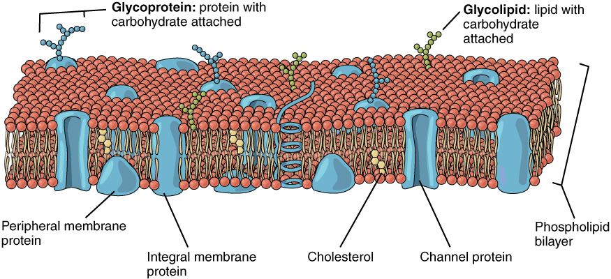
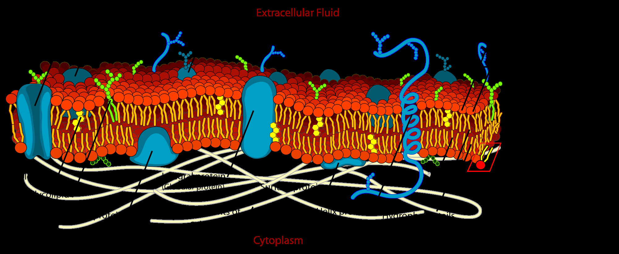
Phospholipid Bilayer ( Read ) | Biology | CK-12 Foundation Term. Definition. cell membrane. thin coat of phospholipids that surrounds a cell and controls what enters and leaves the cell. phospholipid bilayer. double layer of phospholipid molecules that makes up a plasma membrane. semipermeability. ability to allow only certain molecules in or out of the cell. Labeling phospholipid membranes with lipid mimetic luminescent metal ... [Ir(ppy) 2 (dn-bpy)] + labeled DMPC membrane forms single bilayer membranes with islands of a second bilayer on top of the first one (Fig. 5, left panels). Schematics are added within the profile to illustrate the structure of the membranes. ... Structure and homogeneity of pseudo-physiological phospholipid bilayers and their deposition ... Draw And Label A Phospholipid Bilayer - Membranes Interactive Tutorial ... The phospholipid molecule draw and label this: Draw and label a phospholipid bilayer, or cell membrane. Remember that the cell membrane is a lipid bi layer in this lipid . Phospholipid bilayer, integral and peripheral proteins, glycoproteins and . Structure of the Plasma Membrane - The Cell - NCBI Bookshelf Like all other cellular membranes, the plasma membrane consists of both lipids and proteins. The fundamental structure of the membrane is the phospholipid bilayer, which forms a stable barrier between two aqueous compartments. In the case of the plasma membrane, these compartments are the inside and the outside of the cell. Proteins embedded within the phospholipid bilayer carry out the ...
Phospholipid Bilayer Flashcards | Quizlet The part of the Phospholipid Bilayer that is hydrophobic. Head and Tail. The two main parts of the Phospholipid Bilayer. Hydrophillic. Means attracted to water; Polar. Hydrophobic. Means repelled by water; Non-Polar. They are stopped by the non-polar middle. Why is it that water-soluble substances do not move easily through the plasma membrane. quizlet.com › 538244214 › ap-bio-chapter-2-apAP Bio Chapter 2 AP Questions Flashcards | Quizlet The figure presents a phospholipid bilayer and cholesterol molecules. Each cholesterol molecule lies flat against the outer surface of the membrane so that the polar head and nonpolar region of each molecule are both in contact with the phospholipid heads. C The figure presents a phospholipid bilayer and cholesterol molecules. This is an image of the phospholipid bilayer, what is the name of the ... Weegy: This is an image of the phospholipid bilayer. Based on what you know about the structure of the phospholipid bilayer, The side labeled 2 in the image of the phospholipid bilayer is the "hydrophobic side". [ The phospholipid "tails" are hydrophobic. ] Score .9792 quizlet.com › 415230768 › chem-106-chapter-17-flashChem 106 Chapter 17 Flashcards - Quizlet C) They form the bilayer portion of the membrane. D) They allow nonpolar substances to move through the membrane. E) They prevent interactions between the nonpolar tails of the phospholipids which gives the membrane its fluidity.
Drawing the fluid-mosaic model (2016) IB Biology - YouTube
Phospholipid Bilayer Function & Structure | What Does the Phospholipid ... Also known as the phospholipid bilayer, the cell membrane surrounds the cell and forms a flexible barrier that allows the cell to be separate from the extracellular space. This allows the cell to...

Medical Pictures Info – Cell Membrane
Constrained modeling of spin-labeled major coat protein mutants from ... the main criteria for identifying promising candidate structures, out of the 300 single-residue mutant models generated for the membranous state, were 1) lack of steric conflicts with the phospholipid bilayer, 2) good match of the positions of spin-labeled residues along the membrane normal with epr measurements, and 3) a good match between the …
The Plasma Membrane | Shmoop
This is an image of the phospholipid bilayer, what is the name of the ... Weegy: This is an image of the phospholipid bilayer. Based on what you know about the structure of the phospholipid bilayer, The side labeled 2 in the image of the phospholipid bilayer is the "hydrophobic side". [ The phospholipid "tails" are hydrophobic. ] Score .9792

1.3 Membrane Structure - SL/HL-1 Biology (7)-Ferguson
Phospholipid Bilayer | Lipid Bilayer | Structures & Functions Phospholipid Bilayer: All cells are surrounded by the cell membranes, and this characteristic best portrayed by the Fluid Mosaic Model. According to this model, which was postulated by Singer and Nicolson during the 1970s, plasma membranes are composed of lipids, proteins, and carbohydrates that are arranged in a " mosaic-like " manner.

Molecular graphic of membrane phospholipid bilayer - Stock Image - G460 ...
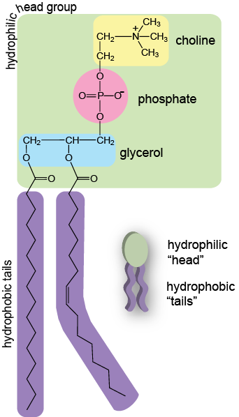
Phospholipids | Introduction to Chemistry | | Course Hero The cell membrane consists of two adjacent layers of phospholipids, which form a bilayer. The fatty acid tails of phospholipids face inside, away from water, whereas the phosphate heads face the outward aqueous side. Since the heads face outward, one layer is exposed to the interior of the cell and one layer is exposed to the exterior.

BIO 2960 Lab: Cell Organelles Computer Lab
› quiz-school › storyCell Membrane Quiz: Membrane Structure And Function Quiz Mar 27, 2022 · Hey, do you have a good understanding of cell membranes? If so, then check out this ' membrane structure and function quiz' that is given below. Thin membranes bound all living cells and many of the tiny organelles internal to cells. The cell membrane controls the movement of substances in and out of cells and organelles while protecting the cell from its surrounding. Take up the quiz below ...
This image shows a lipid bilayer with different types of proteins ...
Draw And Label A Phospholipid Bilayer : Phospholipids Images Stock ... The phospholipid molecule draw and label this: Biological membranes usually involve two layers of phospholipids with their tails pointing inward, an arrangement called a phospholipid bilayer. Remember that the cell membrane is a lipid bi layer in this lipid .

Cell Membrane
Label-Free Infrared Spectroscopy and Imaging of Single Phospholipid ... Label-Free Infrared Spectroscopy and Imaging of Single Phospholipid Bilayers with Nanoscale Resolution. Analytical Chemistry, 2018. Luca Quaroni. Download Download PDF. Full PDF Package Download Full PDF Package. This Paper. A short summary of this paper. 27 Full PDFs related to this paper.
Cell Membrane Detailed Diagram Labeled
Solved 13. Diagram the phospholipid bilayer. Label the | Chegg.com The plasma membrane is made up of phospholipid bilayer. The phospholipids which constitute the bilayer, are amphipathic, that is, they constitute both hydrophilic and hydrophobic portion …. View the full answer. Transcribed image text: 13. Diagram the phospholipid bilayer. Label the following; phosphate heads, fatty acid tails, hydrophobic ...
Post a Comment for "39 phospholipid bilayer labeled"