45 microscope diagram labeled parts
Parts and Function of a Microscope Worksheets - DSoftSchools admin January 4, 2021. Some of the worksheets below are Parts and Function of a Microscope Worksheets with colorful charts and diagrams to help students familiarize with the parts of the microscope along with several important questions and activities with answers. Basic Instructions. Once you find your worksheet (s), you can either click on ... Microscope Parts and Functions With Labeled Diagram and ... Before exploring microscope parts and functions, you should probably understand that the compound light microscope is more complicated than just a microscope with more than one lens. First, the purpose of a microscope is to magnify a small object or to magnify the fine details of a larger object in order to examine minute specimens that cannot ...
Dog Skeleton Anatomy with Labeled Diagram Dec 31, 2021 · Here, in the dog skeleton labeled diagram, I tried to show you the different segments of the forelimb, hindlimb with their bones. Again, I tried to show you all the bones from the vertebrae column of a dog skeleton. In addition, in the diagram, you will find a few identified skull bones. The sternum and the ribs are also identified in the dog ...

Microscope diagram labeled parts
Microscope Types (with labeled diagrams) and Functions Simple microscope labeled diagram Simple microscope functions It is used in industrial applications like: Watchmakers to assemble watches Cloth industry to count the number of threads or fibers in a cloth Jewelers to examine the finer parts of jewelry Miniature artists to examine and build their work Also used to inspect finer details on products Neuron under Microscope with Labeled Diagram - AnatomyLearner But, first, let's try to identify the following features from a neuron with the help of a labelled diagram. Cell body or perikaryon of a neuron Nucleus, cytoplasm, the plasma membrane of a neuron Nissl bodies in the cell body of a neuron An initial segment of axon and axon hillock Dendrites and axons of a neuron Axolemma and myelin sheath PDF Parts of a Microscope Printables - Homeschool Creations Parts of a eyepiece arm stageclips nosepiece focusing knobs illuminator stage objective lenses head base Label the parts of the microscope. You can use the word bank below to fill in the blanks or cut and paste the words at the bottom. Microscope Created by Jolanthe @ HomeschoolCreations.net eyepiece head objective lenses arm focusing knob base ...
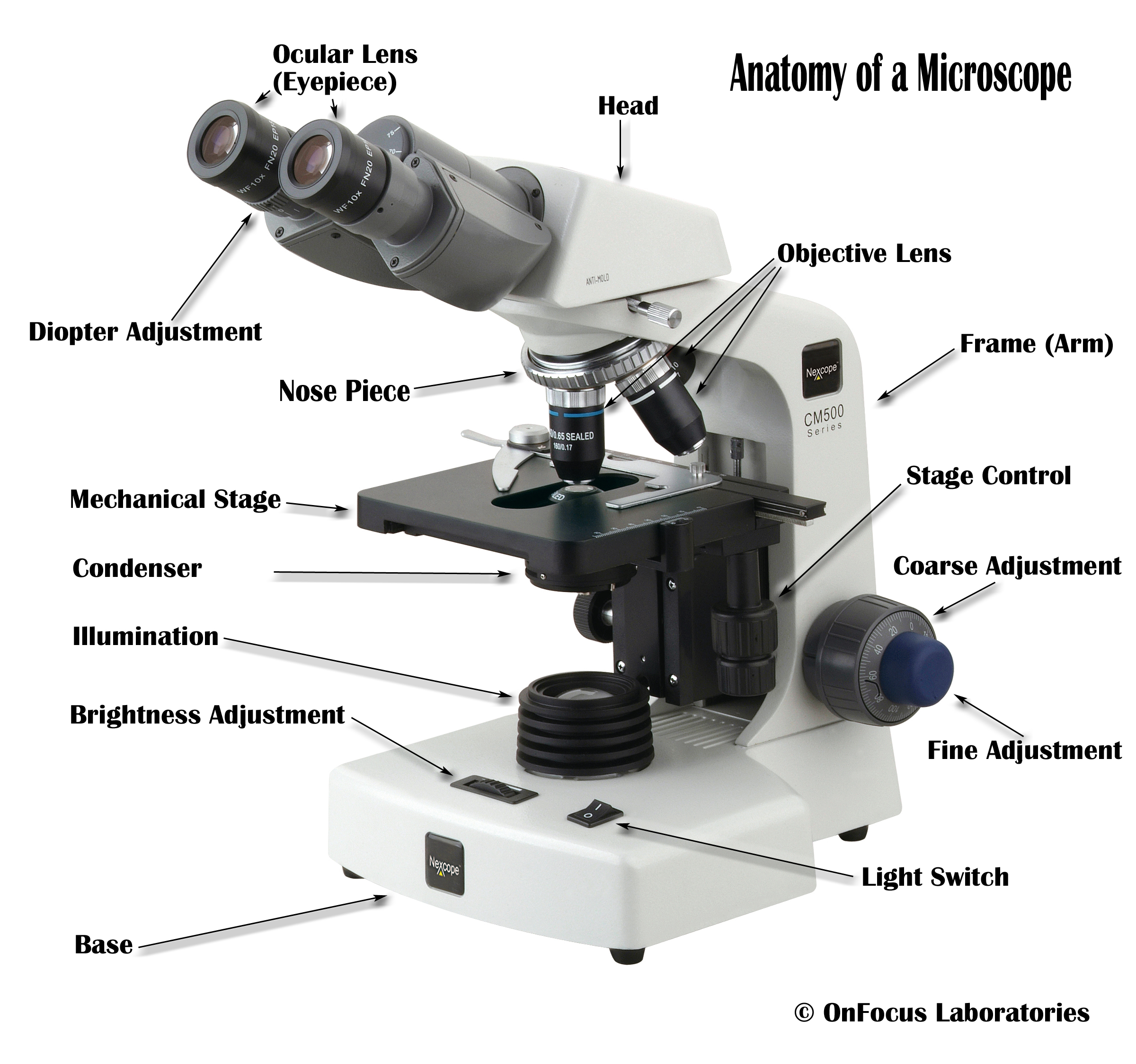
Microscope diagram labeled parts. Cat Skeleton Anatomy with Labeled Diagram - AnatomyLearner May 29, 2021 · Cat skeleton anatomy labeled diagram. Now, I will show you all the bones from the cat skeleton with a diagram. If you find any mistakes in this cat anatomy labeled diagram, please let me know. I hope this cat skeletal system anatomy labeled diagram might help you understand and identify all the cat’s bones. Microscope Parts and Functions Microscope Parts and Functions With Labeled Diagram and Functions How does a Compound Microscope Work?. Before exploring microscope parts and functions, you should probably understand that the compound light microscope is more complicated than just a microscope with more than one lens.. First, the purpose of a microscope is to magnify a small object or to … Compound Microscope Parts, Functions, and Labeled Diagram Common compound microscope parts include: Eyepiece (ocular lens) with or without Pointer: The part that is looked through at the top of the compound microscope. Eyepieces typically have a magnification between 5x & 30x. Monocular or Binocular Head: Structural support that holds & connects the eyepieces to the objective lenses. Parts of Stereo Microscope (Dissecting microscope) – labeled diagram ... Labeled part diagram of a stereo microscope Major structural parts of a stereo microscope. There are three major structural parts of a stereo microscope. The viewing Head includes the upper part of the microscope, which houses the most critical optical components, including the eyepiece, objective lens, and light source of the microscope.
Interactive Bacteria Cell Model - CELLS alive Periplasmic Space: This cellular compartment is found only in those bacteria that have both an outer membrane and plasma membrane (e.g. Gram negative bacteria).In the space are enzymes and other proteins that help digest and move nutrients into the cell. Cell Wall: Composed of peptidoglycan (polysaccharides + protein), the cell wall maintains the overall shape of a … Microscope Parts, Function, & Labeled Diagram - slidingmotion Microscope parts labeled diagram gives us all the information about its parts and their position in the microscope. Microscope Parts Labeled Diagram The principle of the Microscope gives you an exact reason to use it. It works on the 3 principles. Magnification Resolving Power Numerical Aperture. Parts of Microscope Head Base Arm Eyepiece Lens Parts of a microscope with functions and labeled diagram 19-04-2022 · Figure: Diagram of parts of a microscope. There are three structural parts of the microscope i.e. head, base, and arm. Head – This is also known as the body. It carries the optical parts in the upper part of the microscope. Base – It acts as microscopes support. It also carries microscopic illuminators. Simple Microscope - Parts, Functions, Diagram and Labelling Parts of the optical parts are as follows: Mirror - A simple microscope has a plano-convex mirror and its primary function is to focus the surrounding light on the object being examined. Lens - The biconvex lens is placed above the stage and its function is to magnify the size of the object being examined.
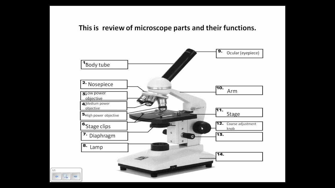
Parts of the Microscope with Labeling (also Free Printouts) Parts of the Microscope with Labeling (also Free Printouts) A microscope is one of the invaluable tools in the laboratory setting. It is used to observe things that cannot be seen by the naked eye. Table of Contents 1. Eyepiece 2. Body tube/Head 3. Turret/Nose piece 4. Objective lenses 5. Knobs (fine and coarse) 6. Stage and stage clips 7. Aperture Labeled Microscope and Basics of Life Diagram | Quizlet Eyepiece the lens at the top that you look through. They are usually 10X or 15X power. Base The bottom of the microscope, used for support and carrying Body tube Connects the eyepiece to the objective lenses Coarse adjustment Brings the specimen into general focus. Fine adjustment Moves the stage slightly to sharpen the image Objective lens Parts of a Compound Microscope and Their Functions Compound microscope mechanical parts (Microscope Diagram: 2) include base or foot, pillar, arm, inclination joint, stage, clips, diaphragm, body tube, nose piece, coarse adjustment knob and fine adjustment knob. Base: It's the horseshoe-shaped base structure of microscope. All of the other components of the compound microscope are supported ... Microscope, Microscope Parts, Labeled Diagram, and Functions Jan 19, 2022 · Illuminator: Illuminator is the most important microscope parts and it serve as light source for a microscope during slide specimen visualization. It is a continuous source of light (110 volts) used in place of a mirror. The mirror of microscope is used to reflect light from the external light source up through the bottom of the stage.
.jpg)
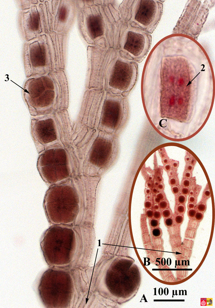
Histology Pictures
Microscope, Microscope Parts, Labeled Diagram, and Functions 19-01-2022 · Revolving Nosepiece or Turret: Turret is the part of the microscope that holds two or multiple objective lenses and helps to rotate objective lenses and also helps to easily change power. Objective Lenses: Three are 3 or 4 objective lenses on a microscope. The objective lenses almost always consist of 4x, 10x, 40x and 100x powers. The most common eyepiece …
Microscope Review.wmv - YouTube
Parts of a Simple Microscope - Labeled (with diagrams) A simple microscope is a very first type of microscope ever created. It consists of simple parts and performs simple functions. Although there are now many advanced microscope types, some applications may still demand the use of a simple microscope. In this article, we are going to discuss the parts and functions of a simple microscope.
Search in gallery
Parts of a microscope with functions and labeled diagram Q. List down the 18 parts of a Microscope. 1. Ocular Lens (Eye Piece) 2. Diopter Adjustment 3. Head 4. Nose Piece 5. Objective Lens 6. Arm (Carrying Handle) 7. Mechanical Stage 8. Stage Clip 9. Aperture 10. Diaphragm 11. Condenser 12. Coarse Adjustment 13. Fine Adjustment 14. Illuminator (Light Source) 15. Stage Controls 16. Base 17.

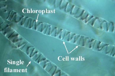
2_Plant_Anatomy
Compound Microscope- Definition, Labeled Diagram, Principle, Parts… 03-04-2022 · Therefore, a microscope can be understood as an instrument to observe tiny elements. The optical microscope often referred to as the light microscope, is a type of microscope that uses visible light and a system of lenses to magnify images of small subjects. There are two basic types of optical microscopes: Simple microscopes; Compound microscopes
Microscope Diagram to Print
Label The Microscope Parts! Diagram | Quizlet Start studying Label The Microscope Parts!. Learn vocabulary, terms, and more with flashcards, games, and other study tools.
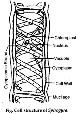
Write in brief the structure of Spirogyra cell - CBSE Class 11 Biology ...
Compound Microscope- Definition, Labeled Diagram, Principle ... Apr 03, 2022 · Therefore, a microscope can be understood as an instrument to observe tiny elements. The optical microscope often referred to as the light microscope, is a type of microscope that uses visible light and a system of lenses to magnify images of small subjects. There are two basic types of optical microscopes: Simple microscopes; Compound microscopes
Print Structure and Function of Plants (Lab Practical #1) flashcards ...
Compound Microscope Parts - Labeled Diagram and their Functions - Rs ... Labeled diagram of a compound microscope Major structural parts of a compound microscope Optical components of a compound microscope Eyepiece Eyepiece tube Objective lenses Nosepiece Specimen stage Coarse and fine focus knobs Rack stop Illuminator Condenser Abbe condenser Iris Diaphragm Condenser Focus Knob Summary An overview of microscopes

Leaf Structure Lab - YouTube
Simple Squamous Epithelium under a Microscope with a Labeled Diagram Histological features of lung parenchyma with microscopic slide images and labeled diagrams. The lung's alveoli give the honeycomb appearance in the parenchyma and lines by flattened simple squamous epithelium. These alveoli are thin-walled and fills with air. From the lung parenchyma labeled diagram, you might identify the following ...
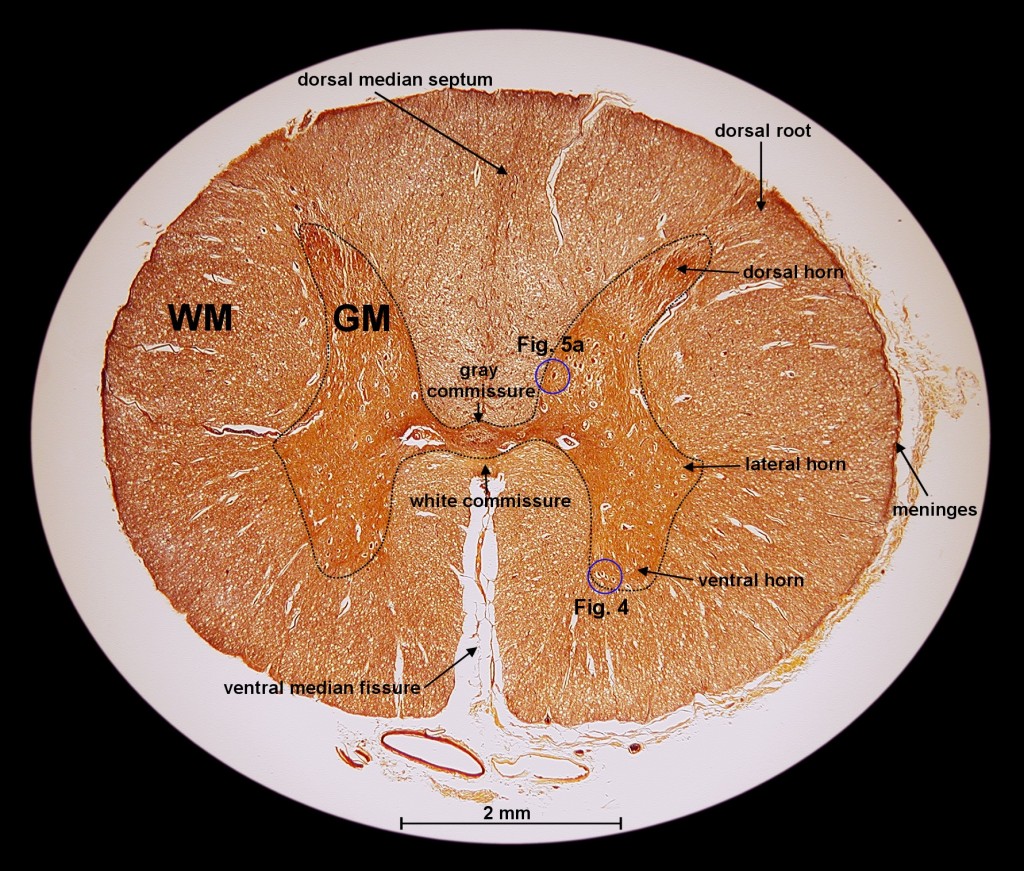
Exploration of the Human Spinal Cord
Label the microscope — Science Learning Hub All microscopes share features in common. In this interactive, you can label the different parts of a microscope. Use this with the Microscope parts activity to help students identify and label the main parts of a microscope and then describe their functions. Drag and drop the text labels onto the microscope diagram.
Post a Comment for "45 microscope diagram labeled parts"