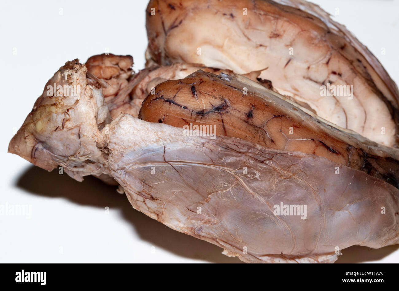
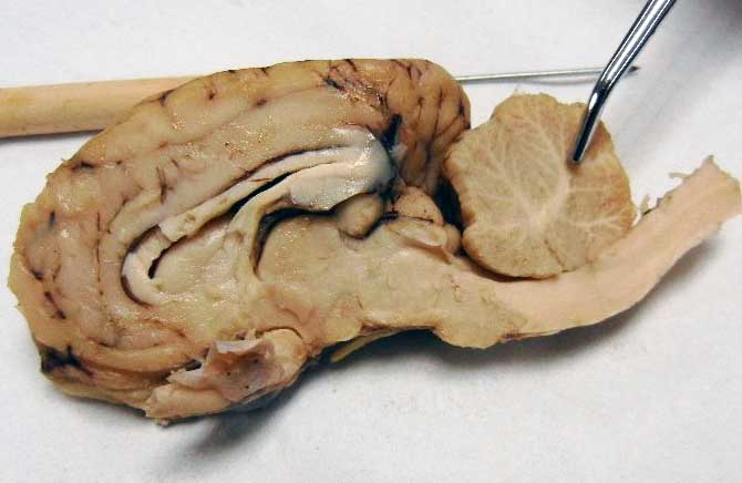
41 sheep brain unlabeled
Sheep brain Flashcards | Quizlet Sheep brain Identification of structures observed during sheep brain dissection. STUDY PLAY dura mater Identify the covering. cerebrum Identify the major brain region. cerebellum Identify the major brain region. olfactory bulb Identify the tip. optic nerve Identify the nerve by name. optic chiasma Identify the "x". optic chiasma Sheep Brain Dissection | Human Anatomy Quiz - Quizizz Play this game to review Human Anatomy. Name this part of the brain. Preview this quiz on Quizizz. Name this part of the brain. Sheep Brain Dissection DRAFT. 6th - 12th grade. 193 times. Biology, Other Sciences. 77% average accuracy. a year ago. mrsturmscience. 0. Save. Edit. Edit. Sheep Brain Dissection DRAFT. a year ago. by mrsturmscience ...
Sheep Brain Label | Dissection, Human brain diagram, Brain anatomy Sheep Brain Label A drawing of the brain with the parts unlabeled. Students can practice naming the parts of the brain, then check their answers with the provided key. Biologycorner 17k followers More information unlabeled brain Find this Pin and more on A&P by Dijana Kovacevic. Human Brain Diagram Brain Gym For Kids Brain Anatomy And Function

Sheep brain unlabeled
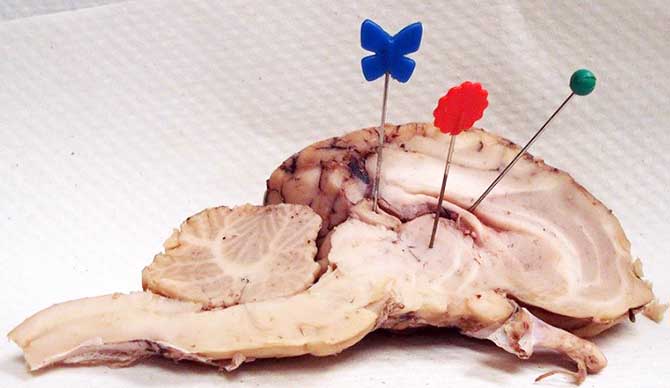
PDF Sheep Brain Midsagittal Section - drcroes.com 5 3 11 6 22 16 18 1. Gray Matter 2. White Matter 3. Corpus Callosum 4. Lateral Ventricle 5. Caudate Nucleus 6. Septum Pellucidum 7. Fornix 8. internal parts of the brain heart diagram anatomy labeled worksheet physiology animals labelled answers human worksheets system label parts structure simple blood wikieducator body unlabeled. Sheep Brain #2 . sheep brain anatomy label nervous system dissection lateral ventricle section sagittal sdmesa classroom edu cerebral diagram aqueduct lesson ... en.wikipedia.org › wiki › Pluto_(Disney)Pluto (Disney) - Wikipedia Curiously enough, however, Pluto was the only standard Disney character not included when the whole gang was reunited for the 1983 featurette Mickey's Christmas Carol, although he did return in The Prince and the Pauper (1990) and Runaway Brain (1995). He also had a cameo at the ending of Who Framed Roger Rabbit (1988).
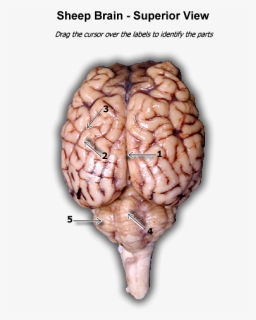
Sheep brain unlabeled. Lab 9—Sheep Brain—Labeled The Sheep's Brain Return to: The Unlabeled Brains Lab 9 Page BIO 137 Main Page Be sure to practice identifying the structures using the unlabeled photos. This page created and maintained by Udo M. Savalli. Last updated August 13, 2005. ... Sheep Brain Neuroanatomy Online Self-Test | KPU.ca - Kwantlen ... Sheep Brain Neuroanatomy Online Self-Test Use each diagram as a reference, and selected the correct answer for each lettered structure. You may find it useful to open the diagrams in a separate window to review while answering each question. Dorsal Surface Dorsal Surface A * Occipital Lobe Temporal Lobe Cerebellum Parietal Lobe Dorsal Surface B * Sheep Brain Instructions - University of Scranton random plate selection (also, to test yourself) This button allows you to toggle between labeled and unlabeled images. On many of the images you will see brackets such as the ones below. A bracket of this type is used to designate an area or region of the brain. Brackets, or lines, which end in small circles designate hollow structures. › articles › srep00028Non-specific binding of antibodies in immunohistochemistry ... Jul 01, 2011 · The current protocols for blocking background staining in immunohistochemistry are based on conflicting reports. Background staining is thought to occur as a result of either non-specific antibody ...
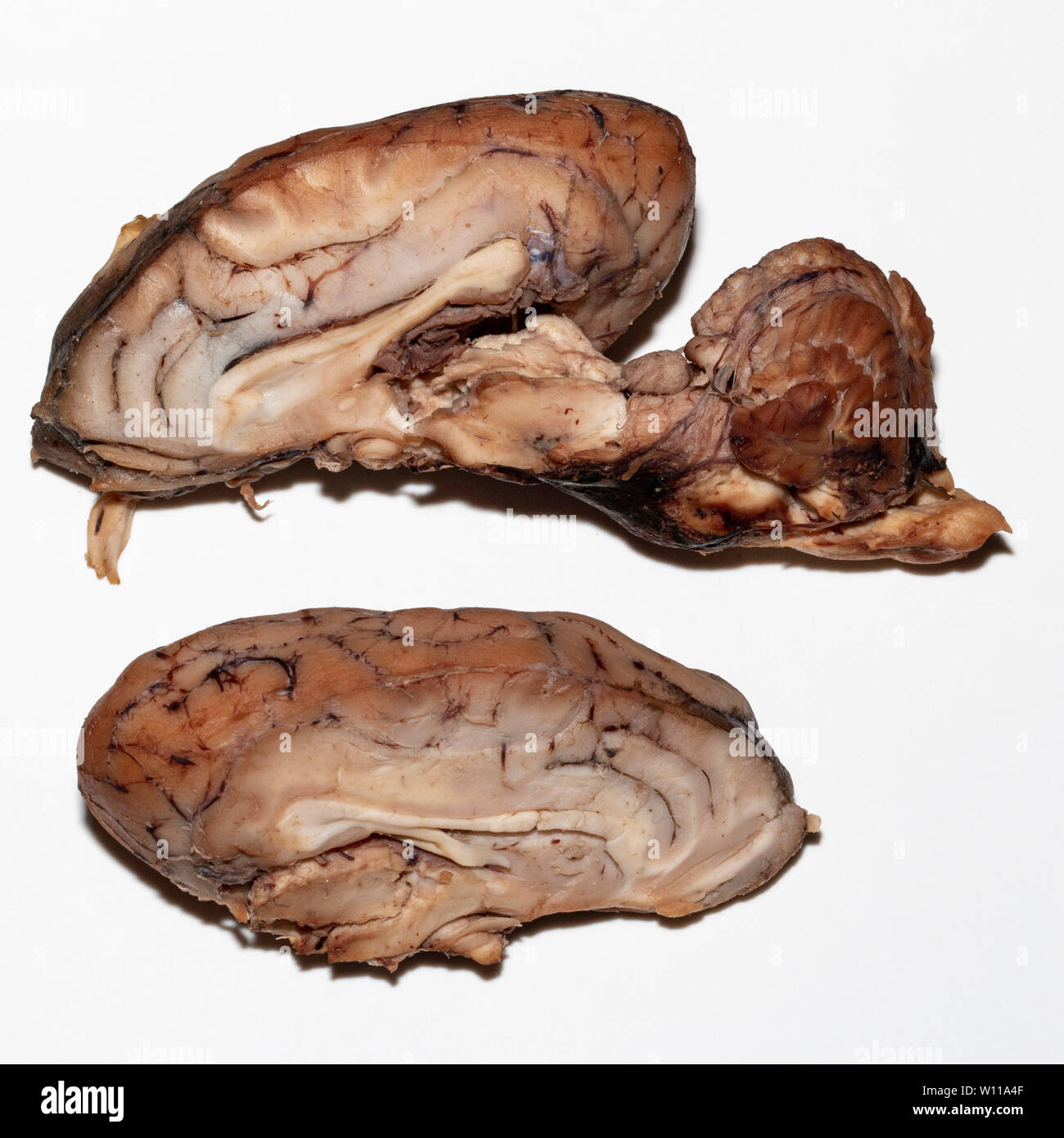
Sheep Brain Dissection labeled Diagram | Quizlet Start studying Sheep Brain Dissection labeled. Learn vocabulary, terms, and more with flashcards, games, and other study tools. Sheep Brain Dissection Project Guide | HST Learning Center Place the brain with the curved top side of the cerebrum facing up. Use a scalpel (or sharp, thin knife) to slice through the brain along the center line, starting at the cerebrum and going down through the cerebellum, spinal cord, medulla, and pons. Separate the two halves of the brain and lay them with the inside facing up. 2. NERVOUS SYSTEM - SHEEP BRAIN IMAGES - San Diego Mesa College Sheep Brain Unlabeled. Sheep Brain Leader-Lined. Sheep Brain Labeled. San Diego Mesa College 7250 Mesa College Drive San Diego, CA 92111-4998 Student Support San Diego Community College District San Diego City College San Diego Mesa College San Diego Miramar College San Diego Continuing Education. › contents › 2921パラフィン包埋または凍結した組織切片 | マウス/ラット脳組織切片 |... パラフィン包埋または凍結したマウス/ラットの脳組織切片をスライドにマウントした製品です。冠状切片14種および矢状断切片,水平切片があります。
› publication › 303806260Machine Learning: Algorithms and Applications - ResearchGate Jul 13, 2016 · rounding a tmosphere; the human brain works to anal yze that information and tak es suitable de cisions acc ordingly . Machines, in con trast, are not intellig ent by nature. › contents › 69162ATCC®の神経細胞株 | ATCC® Neural Cell Lines | フナコシ Sep 24, 2019 · Oliogenglioma/oligoastrocytoma brain tumor stem cell lines (膠原膠腫/乏星細胞腫脳腫瘍幹細胞株) 細胞名をクリックするとメーカーサイトの製品情報,ATCC ® No.クリックすると価格表をご覧いただけます。 Practice Lab Practical on the Sheep Brain - academic.pgcc.edu Identify the cleft labeled 7. Look here for the answer Transverse fissure Identify the shiny membrane visible on the sheep brain surface. Look here for the answer Pia mater In the above picture: Identify the structure labeled 1. Look here for the answer Olfactory bulb Identify the structure labeled 2. Look here for the answer brain of human with label BIO201-Sheep Brain savalli.us brain sheep frontal section labeled bio201 savalli unlabeled return Skull - Bones Of The Cranial Cavity cranial cavity bones skull anatomy resolution poster exploringnature Stafford SammyBear Lakay: Brain Power staffordlakay.blogspot.com
Sheep Brain - Dorsal View The rostral colliculus(large arrow label) and the caudal colliculus(small arrow label) together form the tectumof the midbrain. Also labeled are the pineal body(green), the caudate nucleus(1), the floor of the fourth ventricle(white and pink) and cerebellar peduncles(blue = rostral, red = middle, and yellow = caudal). Go Top
BIO201-Sheep Brain - Savalli This page last updated 18 August 2019 by Udo M. Savalli ()Images and text © Udo M. Savalli. All rights reserved.
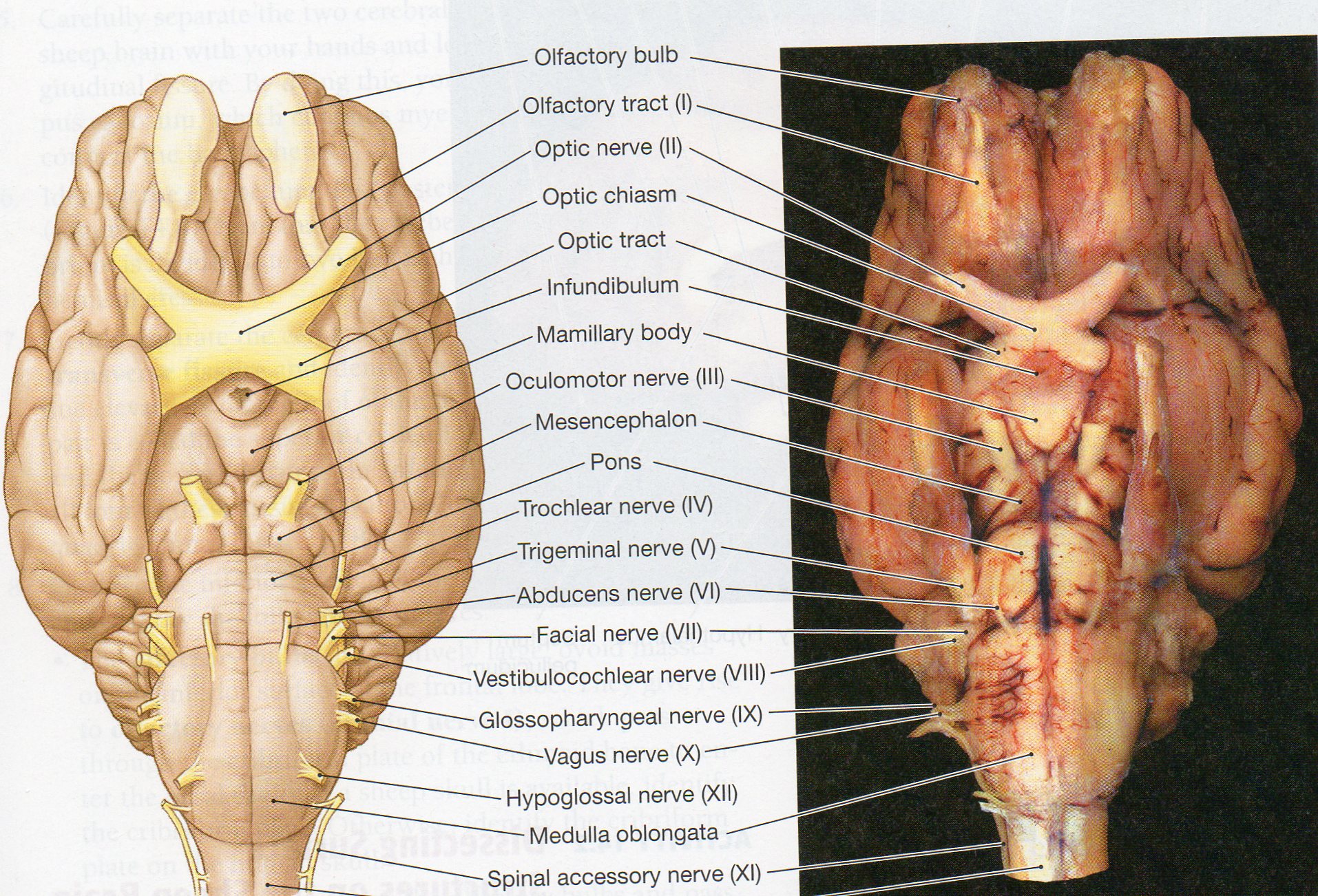
Sheep Brain - Ventral View - University of Minnesota Ventral view of a sheep brain. The optic chiasm (green pic) marks the rostral end of the hypothalamus ( optic nerves are rostral and optic tracts are caudal to the chiasm). Mamillary bodies (red) mark the caudal end of the hypothalamus. Between these, the orange pic is in the lumen of the pituitary stalk (infundibulum).
› CD34-antibody-EP373Y-ab81289CD34重组抗体[EP373Y]_CD34抗体(ab81289)| Abcam中文官网 Rabbit monoclonal IgG (Black) was used as the isotype control, cells without incubation with primary antibody and secondary antibody (Blue) were used as the unlabeled control. Immunohistochemistry (Formalin/PFA-fixed paraffin-embedded sections) - Anti-CD34 antibody [EP373Y] (ab81289)
Unlabeled Sheep Brain Dissection Images and Link (1).pptx Unlabeled Sheep Brain Dissection Images and Link (1).pptx -... School University of Pennsylvania; Course Title BIOL MISC; Uploaded By seperry215yahoo.com. Pages 9 This preview shows page 1 - 2 out of 9 pages. View full document. End of preview. Want to read all 9 pages?
Professor Lapsansky - Western Washington University Photos of the Sheep Brain Dissection are posted below. You may use these photos to help test your recall of sheep brain anatomy for the first lab practical exam. However, not all of the features appear in these pictures. Inferior view of sheep brain - unlabeled; Inferior view of sheep brain - labeled; Mid-sagittal view of sheep brain - unlabeled
Sheep Brain Labeling (part 1) Quiz - By dilatory Sheep Brain Labeling (part 1) Quiz - By dilatory. Science sheep. QUIZ LAB SUBMISSION. Random Science or sheep Quiz.
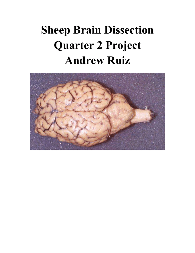
Sheep Brain Dissection with Labeled Images - The Biology Corner 1. The sheep brain is enclosed in a tough outer covering called the dura mater. You can still see some structures on the brain before you remove the dura mater. Take special note of the pituitary gland and the optic chiasma. These two structures will likely be pulled off when you remove the dura mater. Brain with Dura Mater Intact
embryology.med.unsw.edu.au › embryology › indexTestis Development - Embryology - UNSW Sites 2 days ago · Postnatal muscular dystrophy resulting in myotonia, muscle weakness, abnormalities of heart, lungs, eye, brain and endocrine system. There is an associated progressive testicular atrophy (about 80% affected males) leading to Leydig cell hyperproliferation and elevated basal levels of follicle stimulating hormone (FSH). Histology
en.wikipedia.org › wiki › Pluto_(Disney)Pluto (Disney) - Wikipedia Curiously enough, however, Pluto was the only standard Disney character not included when the whole gang was reunited for the 1983 featurette Mickey's Christmas Carol, although he did return in The Prince and the Pauper (1990) and Runaway Brain (1995). He also had a cameo at the ending of Who Framed Roger Rabbit (1988).
internal parts of the brain heart diagram anatomy labeled worksheet physiology animals labelled answers human worksheets system label parts structure simple blood wikieducator body unlabeled. Sheep Brain #2 . sheep brain anatomy label nervous system dissection lateral ventricle section sagittal sdmesa classroom edu cerebral diagram aqueduct lesson ...
PDF Sheep Brain Midsagittal Section - drcroes.com 5 3 11 6 22 16 18 1. Gray Matter 2. White Matter 3. Corpus Callosum 4. Lateral Ventricle 5. Caudate Nucleus 6. Septum Pellucidum 7. Fornix 8.













Post a Comment for "41 sheep brain unlabeled"