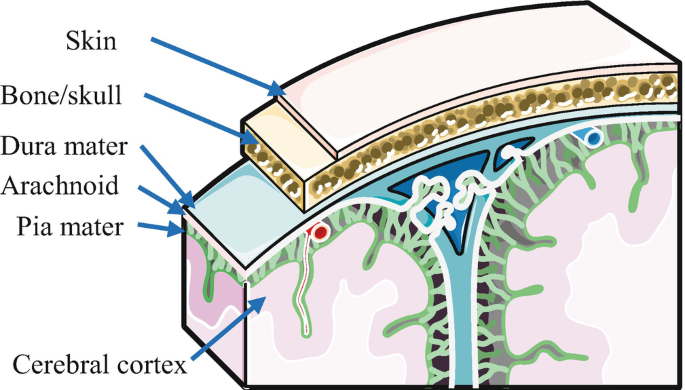
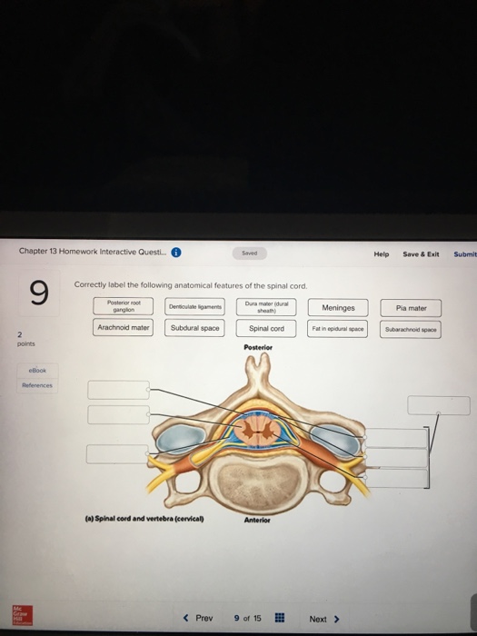
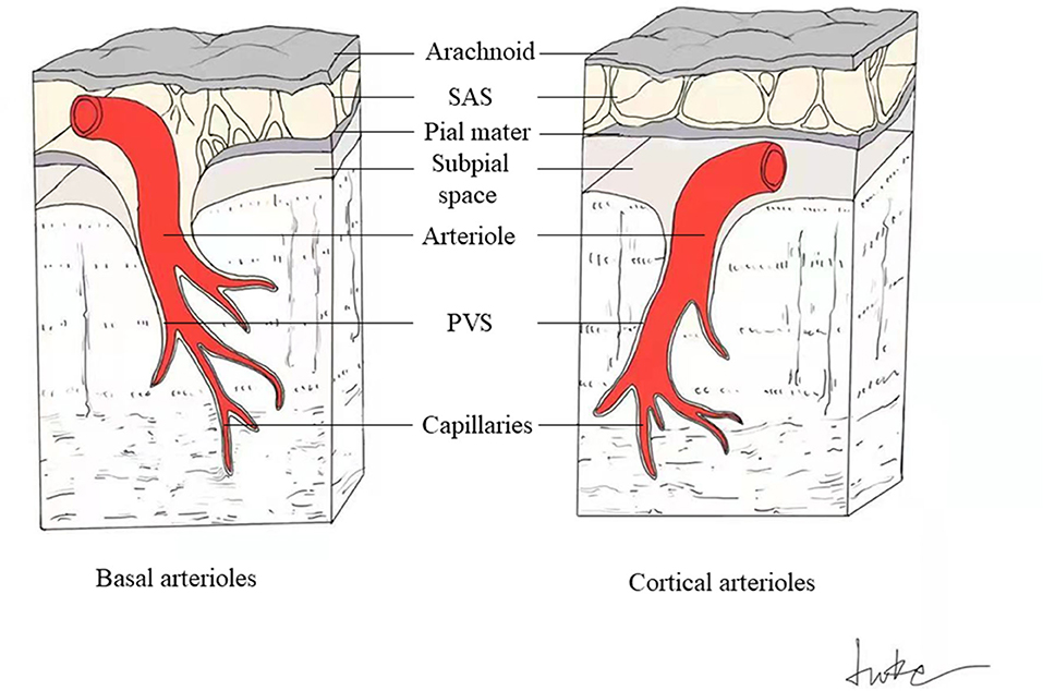
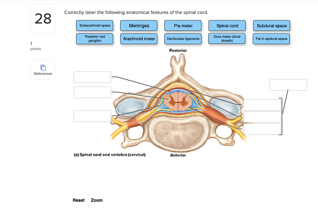
41 correctly label the following meninges and associated structures
Anatomy & Physiology 101 Mastering A&P Ch 13 Practice - Flashcards conduct impulses to smooth and cardiac muscles and glands. Unlock the answer. question. Each of the following effects is associated with stimulation by sympathetic nerves, except. answer. decreased heart rate. Unlock the answer. question. Stimulation of the reticular formation region of the midbrain results in. Anatomy Articles - dummies Nucleic acids These long molecules, found primarily in the cell's nucleus, act as the body's genetic blueprint. They're comprised of smaller building blocks called nucleotides. Each nucleotide, in turn, is composed of a five-carbon sugar (deoxyribose or ribose), a phosphate group, and a nitrogenous base.
Correctly Label the Following Anatomical Features of a Nerve. - Blogger The spinal cord is wrapped in a threelater protective covering called the meninges. Correctly label the following anatomical features of a neuron. Free nerve endings subcutaneous layer oil gland dermis epidermis sweat gland sensory receptor. The optic nerve is the part of the eye that sends electrical signals from the eye to the brain.

Correctly label the following meninges and associated structures
Label the landmarks of the skull in the figure below 1 - Closed (simple) - bone breaks but remains within the skin. 2 - Open (compound) - bone breaks, but part of the bone shaft breaks out of the skin. 3 - greenstick - bone bends and breaks, but not all the way across. 4 - comminuted - bone is broken into more than two segments. 5 - impacted - one end of the broken bone shaft is pushed inside the. The Layers of the Meninges: Medical Vocabulary - Study.com The meninges are three fibrous membranes that enclose the central nervous system, which consists of the brain and spinal cord. The thick and tough outermost meningeal layer is called the dura mater . Positions and Functions of the Four Brain Lobes | MD-Health.com The occipital lobe, the smallest of the four lobes of the brain, is located near the posterior region of the cerebral cortex, near the back of the skull. The occipital lobe is the primary visual processing center of the brain. Here are some other functions of the occipital lobe: Visual-spatial processing. Movement and color recognition.
Correctly label the following meninges and associated structures. Anatomical Planes of Body | What Are They?, Types & Position In Body located anteriorly to the dorsal cavity and houses the space inside the skull. This cranial cavity is occupied with the brain, meninges, and cerebrospinal fluid. Ventral Cavity This cavity is located interiorly in front of the body and a house of many different organ systems. Besides, this cavity is divided by the diaphragm into anterior and Spinal cord: Anatomy, structure, tracts and function | Kenhub The spinal cord and spinal nerve roots are wrapped within three layers called meninges. The outermost is the dura mater, underneath it is the arachnoid mater, and the deepest is the pia mater. Dura mater has two layers (periosteal and meningeal), between which is the epidural space. Label the landmarks of the skull in the figure below The cavities, or spaces, of the body contain the internal organs, or viscera. The two main cavities are called the ventral and dorsal cavities. The ventral is the larger cavity and is subdivided into two parts (thoracic and abdominopelvic cavities) by the diaphragm, a dome-shaped respiratory muscle.. Thoracic cavity. baptismal vows sda pdf Central nervous system: Anatomy, structure, function | Kenhub They are enveloped and protected by three layers of meninges, and encased within two bony structures; the skull and vertebral column, respectively. The brain consists of the cerebrum, subcortical structures, brainstem and cerebellum. The spinal cord continues inferiorly from the brainstem and extends through the vertebral canal.
Anatomy And Physiology Archive | February 17, 2022 - Chegg A) increase the volts per division B decrease the volts per division C decrease the milliseconds per division D 1 answer For the following questions, use these values: Ion Inside Outside Na+ 15 150 K+ 150 5 C1- 110 10 Ca++ 0.0001 2.5 Calculate the Nernst potential for Ca++ Calculate the Nernst potential for Na+ 1 answer 7 Meningitis Nursing Care Plans - Nurseslabs Meningitis is the inflammation of the meninges of the brain and spinal cord as a result of either bacteria, viral or fungal infection.Bacterial infections may be caused by Haemophilus influenzae type b, Neisseria meningitidis (meningococcal meningitis), and Streptococcus pneumoniae (pneumococcal meningitis). Those at greatest risk for this disease are infants between 6 and 12 months of age ... What Is the Central Nervous System (CNS)? - Verywell Mind Dura mater: From the Latin words meaning "hard mother," this is the top layer of the meninges found directly under the bones of the skull and vertebrae.It is composed of dense connective tissue. Arachnoid mater: The second layer of the meninges is a spider-like, transparent membrane made up of collagen and elastic fibers.; Pia mater: From the Latin for "soft mother," this protective layer is ... (Get Answer) - Correctly label the following anatomical features of the ... Correctly label the following anatomical features of the spinal cord. Fat in epidural space Subdural space Spinal nerve Dura mater (dural sheath) Vertebral body Posterior root ganglion Arachnoid mater Spinous process Posterior Spinous process Fat in epidural space Vertebral body (a) Spinal cord and vertebra (cervical) Anterior Apr 11 2022 05:44 AM
cisco aci software download - lineartdrawingsanimesimple The Data Center Networking Software Subscription suites bring customer flexibility when migrating to the software-defined Cisco policy-driven management and. Step 2 Extract the tools-msc- targz file to the directory from which you. Others help us improve services and the user experience or to. S2.B1 Meninges - ProProfs Quiz A. Predisposes the patient to infection, because bacteria can easily get inside. B. Is a developmental defect associated with spina bifida. C. Is where the mesoderm fails to dissociated from neuroectoderm, leaving an epithelium lined channel to the surface of the skin. D. Results in recurrent meningitis. 5. Meninges: Dura, arachnoid, pia, meningeal spaces | Kenhub Meninges of the brain The meninges are the three membranes that envelop the brain and spinal cord and separate them from the walls of their bony cases ( skull and vertebral column ). Based on their location, meninges are referred to as the cranial meninges which envelop the brain, and spinal meninges which envelop the spinal cord. Anatomy And Physiology Archive | April 08, 2022 | Chegg.com Anatomy And Physiology Archive: Questions from April 08, 2022. In the diagram of an artery below, which letter corresponds to: a) tunica adventitia D b) tunica intima A c) vessel lumen B d) tunica media B e) the initial site where LDL infiltrates С f) contains e. 1 answer.
Body Cavities and Membranes: Labeled Diagram, Definitions - EZmed Body Cavity Definition: A space or compartment in the body that houses organs and structures. Dorsal Cavity and Ventral Cavity There are a number of different cavities in the body. To make them easier to understand, let's create a flow chart. First, there are 2 main cavities in the body. They include the dorsal cavity and the ventral cavity.
Spinal Nerves: Anatomy, Function, and Treatment - Verywell Health A total of 31 pairs of spinal nerves control motor, sensory, and other functions. Spinal nerves can be impacted by a variety of medical conditions, resulting in pain, weakness, or decreased sensation. A pinched nerve, which occurs when there is pressure on or compression of a spinal nerve, is a common issue. 1.
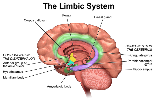
Fornix | Structure, Function, Connections, Role & Summary In this section, we will talk about the structure, histology, connections, and relations of fornix. The word fornix is a Latin word meaning "arch". This name is used because of the arch-shaped structure. The fornix consists of three parts; crus, body, commissure, and anterior columns. We will discuss the details of all these parts below.
Anatomy, Central Nervous System - StatPearls - NCBI Bookshelf The brain is an organ of nervous tissue that is responsible for responses, sensation, movement, emotions, communication, thought processing, and memory. Protection for the human brain comes from the skull, meninges, and cerebrospinal fluids. The nervous tissue is extremely delicate and can suffer damage by the smallest amount of force.
Aseptic Meningitis - StatPearls - NCBI Bookshelf Aseptic meningitis is a term used to define inflammation of the brain linings, called meninges, due to various etiologies with negative cerebrospinal fluid (CSF) bacterial cultures. Many studies and books determine it by showing CSF pleocytosis of more than five cells/mm3.[1] It is one of the most common, usually benign, inflammatory disorders of the meninges.[2]
Meninges, Ventricles, CSF and brain blood supply | Kenhub Meninges. The three membranes that envelop and protect the surfaces of the brain and spinal cord. Meninges are: Dura mater, pia mater and arachnoid mater. Ventricles of the brain. A system of interconnected spaces within the brain through which the cerebrospinal fluid criculates. The ventricles are: the two lateral, third and fourth ventricle.
Frank ICSE Class 10 Biology Solutions Chapter 9 Nervous System ICSE Solutions for Class 10. Question 1. Name the following: (i) Nervous system including brain and spinal cord. (ii) Sympathetic and parasympathetic jointly forms. (iii) Terminal end of spinal cord. (iv) The third neuron that is involved in reflexes, other than simple reflex. (v) Study of structure and functions of nervous system.
Match the Following Structures With the Correct Descriptions. Identify the meningeal or associated structures described here. Involved in twitching motility C. Match the following structures with the primary germ layer from which they were derived. B 19 Smallest particle of an element that retains its properties. Outermost covering of the brain composed of tough fibrous connective tissue 2.
Nervous system: Structure, function and diagram | Kenhub Neurons, or nerve cell, are the main structural and functional units of the nervous system. Every neuron consists of a body (soma) and a number of processes (neurites). The nerve cell body contains the cellular organelles and is where neural impulses ( action potentials) are generated.
Anatomy And Physiology Archive | November 18, 2021 - Chegg Anatomy And Physiology Archive: Questions from November 18, 2021. what muscles are these? 1 answer Alzheimer's disease 1.- Where are memories "stored" in the brain? 2.- Why do some elderly people act as if their memories were "erased"? 3.- Are language areas affected in this disease?
Positions and Functions of the Four Brain Lobes | MD-Health.com The occipital lobe, the smallest of the four lobes of the brain, is located near the posterior region of the cerebral cortex, near the back of the skull. The occipital lobe is the primary visual processing center of the brain. Here are some other functions of the occipital lobe: Visual-spatial processing. Movement and color recognition.
The Layers of the Meninges: Medical Vocabulary - Study.com The meninges are three fibrous membranes that enclose the central nervous system, which consists of the brain and spinal cord. The thick and tough outermost meningeal layer is called the dura mater .
Label the landmarks of the skull in the figure below 1 - Closed (simple) - bone breaks but remains within the skin. 2 - Open (compound) - bone breaks, but part of the bone shaft breaks out of the skin. 3 - greenstick - bone bends and breaks, but not all the way across. 4 - comminuted - bone is broken into more than two segments. 5 - impacted - one end of the broken bone shaft is pushed inside the.



:watermark(/images/watermark_only_sm.png,0,0,0):watermark(/images/logo_url_sm.png,-10,-10,0):format(jpeg)/images/anatomy_term/parasympathetic-nervous-system/NW2chCFVkZmbGDJc7RjXGg_Parasympathetic_nervous_system.jpeg)









Post a Comment for "41 correctly label the following meninges and associated structures"