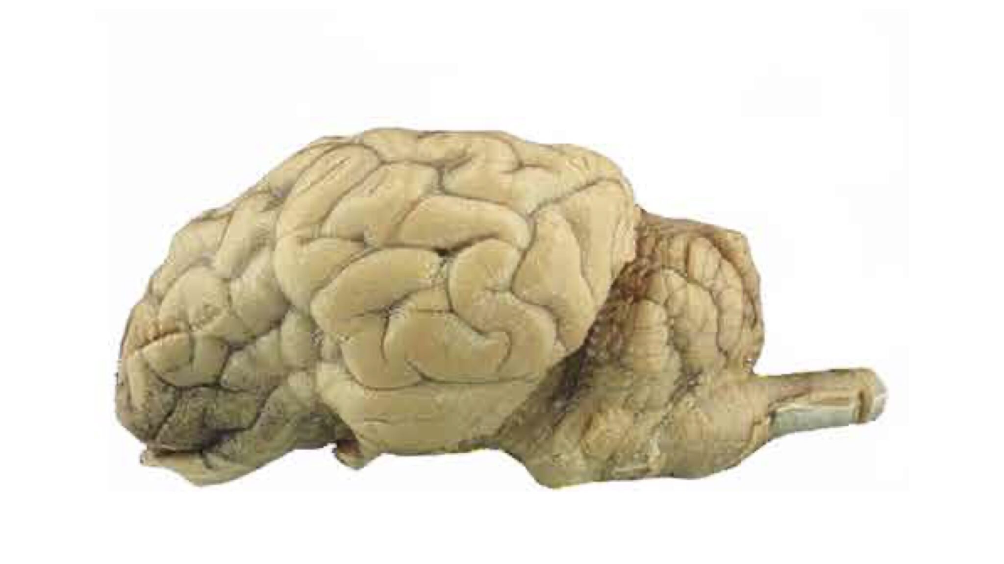
44 unlabeled sheep brain
Unlabeled Sheep Brain Dissection Images and Link (1).pptx Unlabeled Sheep Brain Dissection Images and Link (1).pptx -... School University of Pennsylvania; Course Title BIOL MISC; Uploaded By seperry215yahoo.com. Pages 9 This preview shows page 1 - 2 out of 9 pages. View full document. End of preview. Want to read all 9 pages? Lab 9—Sheep Brain—Labeled - Bluegrass Community and Technical College The Sheep's Brain Return to: The Unlabeled Brains Lab 9 Page BIO 137 Main Page Be sure to practice identifying the structures using the unlabeled photos. This page created and maintained by Udo M. Savalli. Last updated August 13, 2005. ...
Sheep brain Flashcards | Quizlet Only $35.99/year Sheep brain Flashcards Learn Test Match Flashcards Learn Test Match Created by nina_ureke Identification of structures observed during sheep brain dissection. Terms in this set (29) dura mater Identify the covering. cerebrum Identify the major brain region. cerebellum Identify the major brain region. olfactory bulb

Unlabeled sheep brain
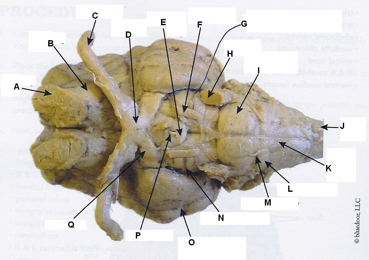
Brain Labeling Worksheet brain anatomy sheep dissection label unlabeled worksheet lab sheet systems body biology labeled guide ap biologycorner parts lateral psychology practice. Sheep Brain Dissection @ Fort Vancouver! - NW NOGGIN: Neuroscience nwnoggin.org. brain sheep dissection superior vancouver fort skyview storm colliculi prepared handout angela johnson nwnoggin Sheep Brain - midsagittal Flashcards | Quizlet Only $2.99/month Sheep Brain - midsagittal STUDY Flashcards Learn Write Spell Test PLAY Match Gravity Created by ryankidd01 Terms in this set (20) frontal lobe Identify the lobe labeled 1 parietal lobe Identify the lobe labeled 2 occipital lobe Identify the lobe labeled 3 arbor vitae Identify the structure labeled 4 spinal cord Sheep Brain - Ventral View - University of Minnesota Ventral view of a sheep brain. The optic chiasm (green pic) marks the rostral end of the hypothalamus ( optic nerves are rostral and optic tracts are caudal to the chiasm). Mamillary bodies (red) mark the caudal end of the hypothalamus. Between these, the orange pic is in the lumen of the pituitary stalk (infundibulum). To return to the dorsal ...
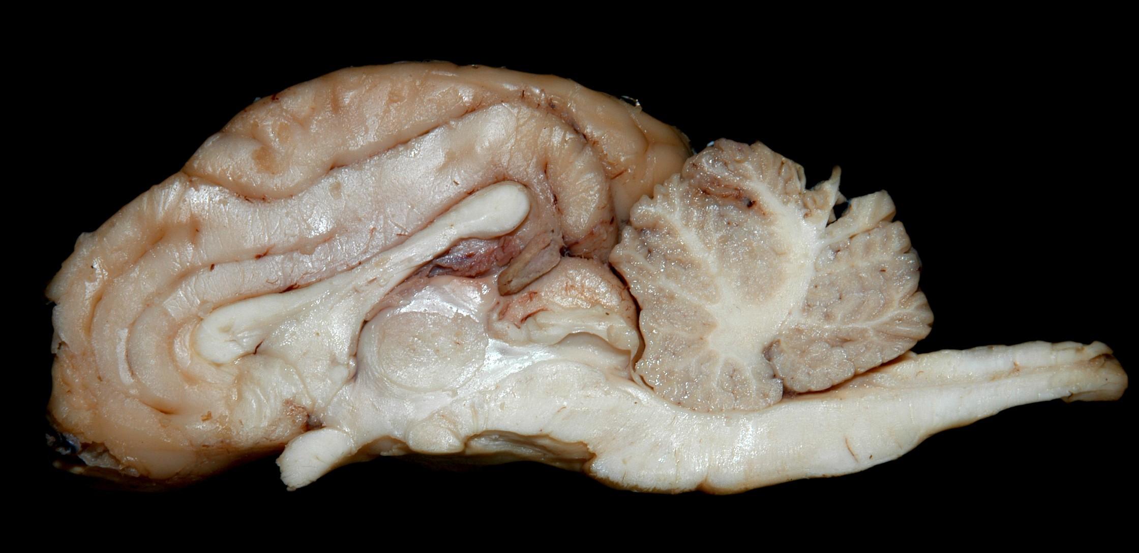
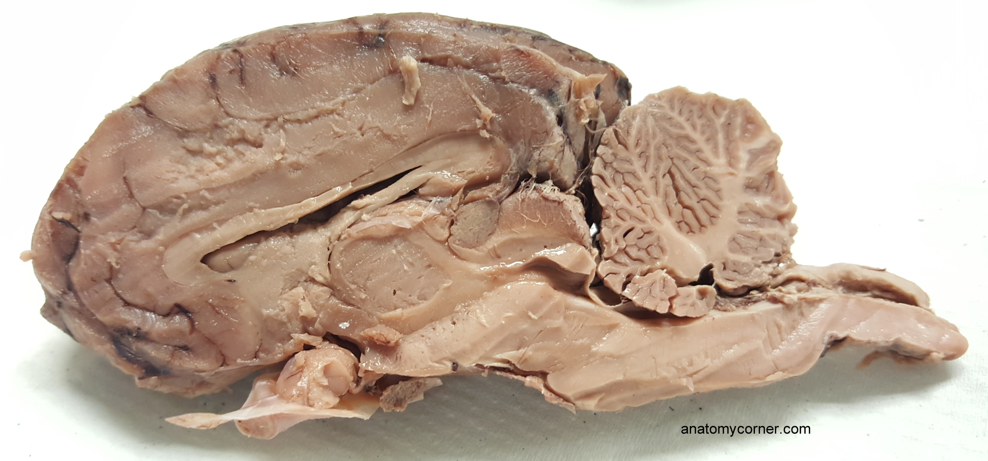
Unlabeled sheep brain. Sheep Brain Dissection Project Guide | HST Learning Center Place the brain with the curved top side of the cerebrum facing up. Use a scalpel (or sharp, thin knife) to slice through the brain along the center line, starting at the cerebrum and going down through the cerebellum, spinal cord, medulla, and pons. Separate the two halves of the brain and lay them with the inside facing up. 2. Sheep Brain Dissection with Labeled Images - The Biology Corner 1. The sheep brain is enclosed in a tough outer covering called the dura mater. You can still see some structures on the brain before you remove the dura mater. Take special note of the pituitary gland and the optic chiasma. These two structures will likely be pulled off when you remove the dura mater. Brain with Dura Mater Intact Sheep Brain Dissection labeled Diagram | Quizlet Start studying Sheep Brain Dissection labeled. Learn vocabulary, terms, and more with flashcards, games, and other study tools. Sheep Brain Neuroanatomy Online Self-Test | KPU.ca - Kwantlen ... Sheep Brain Neuroanatomy Online Self-Test Use each diagram as a reference, and selected the correct answer for each lettered structure. You may find it useful to open the diagrams in a separate window to review while answering each question. Dorsal Surface Dorsal Surface A * Occipital Lobe Temporal Lobe Cerebellum Parietal Lobe Dorsal Surface B *
sheep-brain-labeled-brain-01.jpg - | Course Hero sheep-brain-labeled-brain-01.jpg - School Central Texas College; Course Title BIOLOGY 1406; Uploaded By CountDanger2952. Pages 1 Ratings 100% (1) 1 out of 1 people found this document helpful; ... endocrine-system-unlabeled-diagram.jpg. Indian River State College. BIOLOGY 123. sheep brain labeling worksheet diagram crab brain 2007 worksheet worksheeto unlabeled labeled via sheep. Formulas Poker: This Is A Card Game To Practice Writing Chemical ... sheep brain dissection anatomy lab dorsal superior bi diagrams nervous system companion procedure names quizlet biologyjunction human. Labeled Diagrams of the Human Brain You'll Want to Copy Now The average dimension of the adult human brain is 5.5 inches in width and 6.5 inches in length. The height of the human brain is about 3.6 inches and it weighs about 4 to 5 lbs at birth and 3 lbs in adults. The total surface area of the cerebral cortex is about 2,500 cm2 and when stretched, it will cover the area of a night table. anatomy brain diagram worksheet Sheep brain dissection guide. Neuron diagram unlabeled nervous worksheet system nerve cell impulse neurone function kinds different muscle wikieducator anatomy fibres match kind each ... NAME LAB TIME/DATE Gross, 11 Best Images of Brain Diagram Worksheet - Unlabeled Brain Diagram and also Parts Of Brain And Their Functions Chart - Human Anatomy
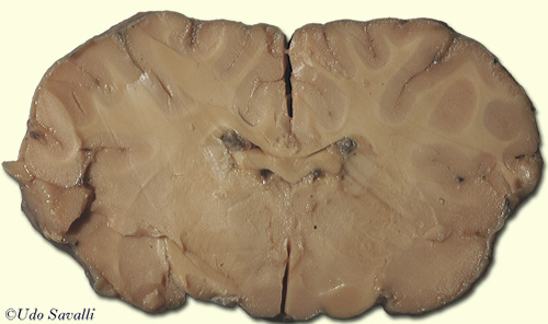
American Express Bio201 Sheep Brain Description : Bio201 ... physiology animals labelled answers human worksheets system label parts structure simple blood wikieducator body unlabeled. Sheep Brain #2 . sheep brain anatomy label nervous system dissection lateral ventricle section sagittal sdmesa classroom edu cerebral diagram aqueduct lesson. BIO201-Sheep Brain - Savalli This page last updated 18 August 2019 by Udo M. Savalli (dr.udo @ savalli.us)Images and text © Udo M. Savalli. All rights reserved. sheep brain labeling worksheet Dissection unlabeled sheep brain labeling worksheet Sheep Brain Dissection Worksheet. 17 Pics about Sheep Brain Dissection Worksheet : 12 Best Images of Brain Parts Worksheet - Brain Label Worksheet, Human, 31 Sheep Brain Dissection Worksheet - Worksheet Resource Plans and also 31 Sheep Brain Dissection Worksheet - Worksheet Resource Plans. PDF Sheep Brain Practical Study Guide - auburn.k12.il.us Sheep Brain Practical Study Guide. Dura Mater. Olfactory Bulb Pituitary Gland Dura Mater Optic Chiasm. Corpus Callosum Longitudinal Fissure Lateral Ventricle Gray Matter White Matter. Arbor Vitae "Tree of Life" Cerebellum "Little Brain" ...
Sheep Brain Dissection Images
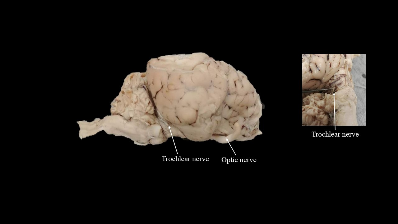
Practice Lab Practical on the Sheep Brain - PGCC Identify the cleft labeled 7. Look here for the answer Transverse fissure Identify the shiny membrane visible on the sheep brain surface. Look here for the answer Pia mater In the above picture: Identify the structure labeled 1. Look here for the answer Olfactory bulb Identify the structure labeled 2. Look here for the answer
SHEEP BRAIN VENTRAL VIEW Flashcards | Chegg.com
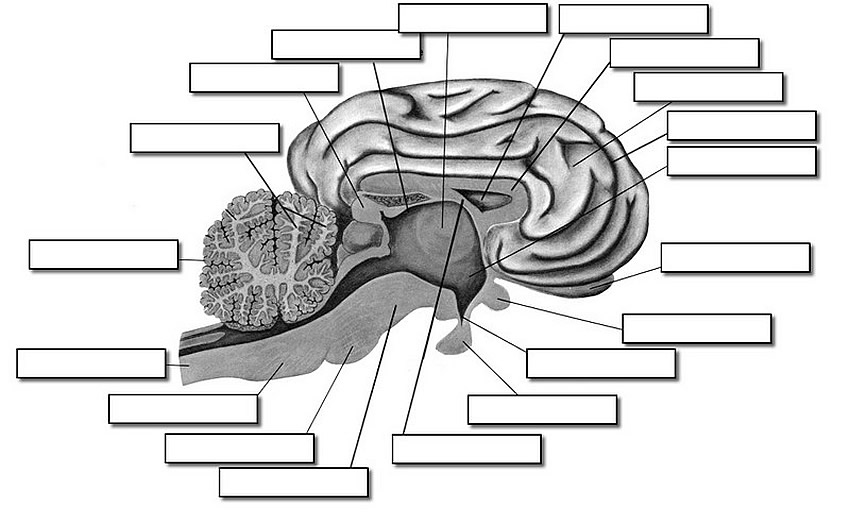
Sheep Brain Label | Dissection, Human brain diagram, Brain anatomy Sheep Brain Label A drawing of the brain with the parts unlabeled. Students can practice naming the parts of the brain, then check their answers with the provided key. Biologycorner 17k followers More information unlabeled brain Find this Pin and more on A&P by Dijana Kovacevic. Nervous System Lesson Nervous System Anatomy Human Brain Diagram
Sheep brain images | Lab | Amherst College
Sheep Brain - Ventral View - University of Minnesota Ventral view of a sheep brain. The optic chiasm (green pic) marks the rostral end of the hypothalamus ( optic nerves are rostral and optic tracts are caudal to the chiasm). Mamillary bodies (red) mark the caudal end of the hypothalamus. Between these, the orange pic is in the lumen of the pituitary stalk (infundibulum). To return to the dorsal ...

sheep brain dissection labeling ex. 14 Diagram | Quizlet
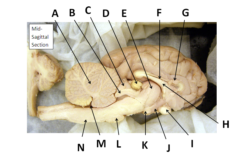
Sheep Brain - midsagittal Flashcards | Quizlet Only $2.99/month Sheep Brain - midsagittal STUDY Flashcards Learn Write Spell Test PLAY Match Gravity Created by ryankidd01 Terms in this set (20) frontal lobe Identify the lobe labeled 1 parietal lobe Identify the lobe labeled 2 occipital lobe Identify the lobe labeled 3 arbor vitae Identify the structure labeled 4 spinal cord

Sheep Brain Dissection Images
Brain Labeling Worksheet brain anatomy sheep dissection label unlabeled worksheet lab sheet systems body biology labeled guide ap biologycorner parts lateral psychology practice. Sheep Brain Dissection @ Fort Vancouver! - NW NOGGIN: Neuroscience nwnoggin.org. brain sheep dissection superior vancouver fort skyview storm colliculi prepared handout angela johnson nwnoggin

Ventral View of Sheep Brain Diagram | Quizlet

Sheep Brain

Sheep Brain Dissection Vita AP Psych by Maria Vita
Sheep Brain
BIO201-Sheep Brain
SCB209 - Lab2 - Natural Sciences Open Educational Resources

Dissecting Sheep Brains With Sixth Graders | Brains Explained
DISSECTION OF THE SHEEP'S BRAIN
Lab: Sheep Brain Dissection
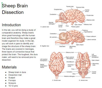
Lab 12 - The Brain and Cranial Nerves
Sheep Neuroanatomy Lab- Labeling Worksheet Figure 1: Dorsal view

Ventral view of a sheep brain Flashcards | Quizlet
Sheep Brain Dissection
3.2: Sheep brain - Medicine LibreTexts
Sheep Brain Flashcards - Easy Notecards

Sheep Brain Dissection | Carolina.com
Sheep brain dissection worksheet
Sheep Brain Dissection Guide

Sheep Brain
Sheep Brain Neuroanatomy Online Self-Test | KPU.ca - Kwantlen ...
Neuroanatomy: Dissection of the Sheep Brain
hey! i want to die - sheep brain lab practical | Conjunto de ...
Sheep Brain Explora on Guide

Sheep Brain Dissection Images
Neuroanatomy: Dissection of the Sheep Brain

brain-sheep-chiasma-1280px | Brain, unlabeled, ventral view ...
Sheep Brain
PhysioEx Exercise 3: Neurophysiology

Sheep Brain

BIO201-Sheep Brain
Lab - Sheep Brain: MAH-Summer 2019-Anatomy and Physiology I
BRAIN ANATOMY The anatomy of the brain is often discussed in ...

Welcome to the sheep brain structure review! arachnoid layer ...

Sheep Brain Quiz

Carolina's Perfect Solution Sheep Brain, Dura Mater Intact, Plain, Pail
Dissection of the Sheep Brain

Lab Diary for Sheep Brain Dissection - Behavioral Neuro Lab mdp
Lab: Sheep Brain Dissection
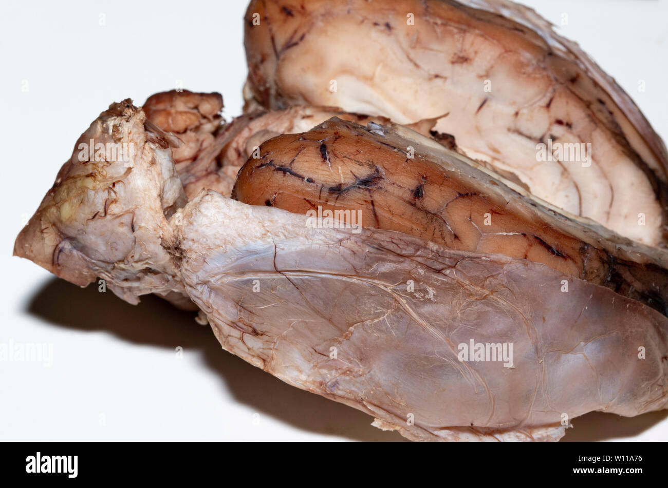
Close up of a sheep brain showing the Meninges Stock Photo ...
Post a Comment for "44 unlabeled sheep brain"