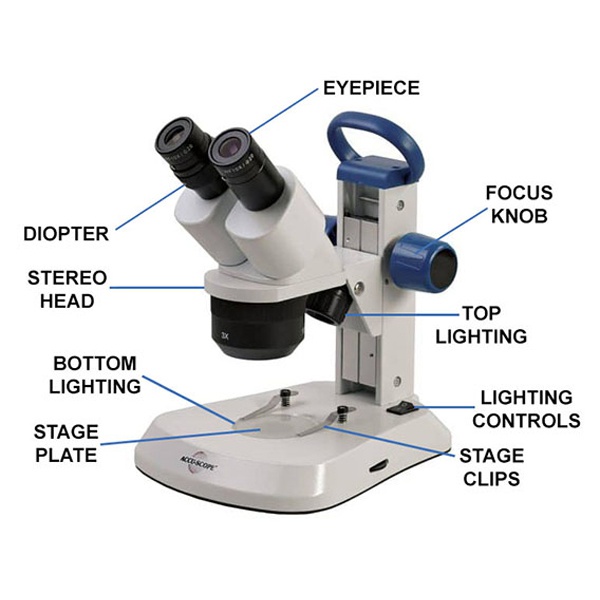
40 draw and label a binocular microscope
Join LiveJournal Password requirements: 6 to 30 characters long; ASCII characters only (characters found on a standard US keyboard); must contain at least 4 different symbols; Microscope Drawing: How to Sketch Microscope Slides How to Draw Microscope Slides Organize and orient your field of view: To begin, draw a circle as large as possible with a pencil. An 8.5 x 11-inch piece of paper is good size for beginners. The circle represents what you see through the eyepiece of the microscope. Using thin lines, divide the circle into quarters in order to organize the picture.
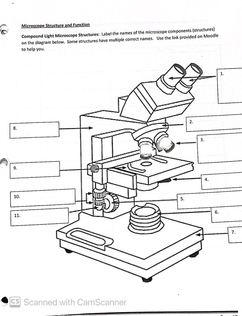
Labeling A Microscope AnswersThis printable worksheet of … 10th : 8 Best Images of Using A Microscope Worksheet - Compound Microscope, Pin on School 2015 and also 27 Microscope Worksheet Answer Key - Worksheet Resource Plans. Arms – This is the part connecting the base and to the head and the eyepiece tube to the base of the microscope. 10000+ results for 'label a microscope'. Label the Microscope Quiz.

Draw and label a binocular microscope
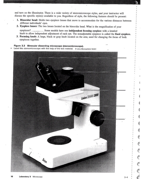
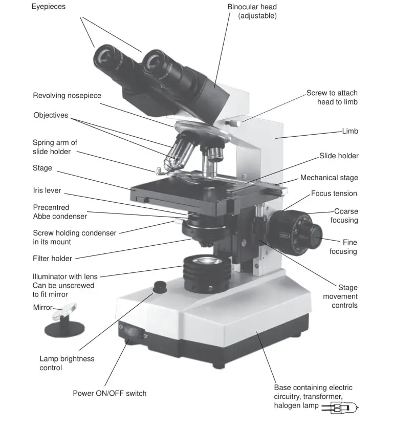
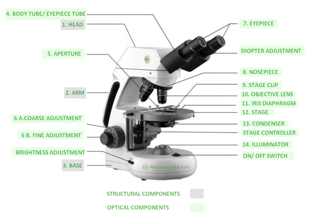
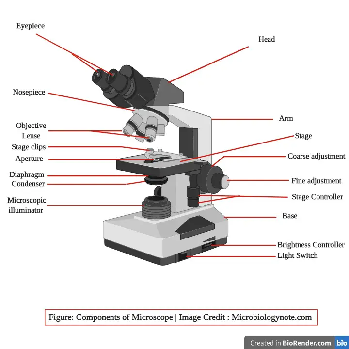
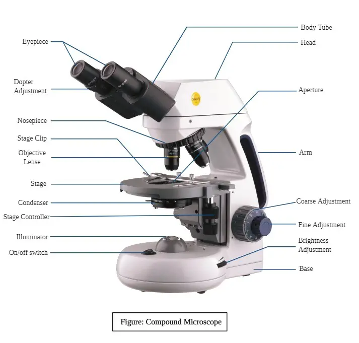
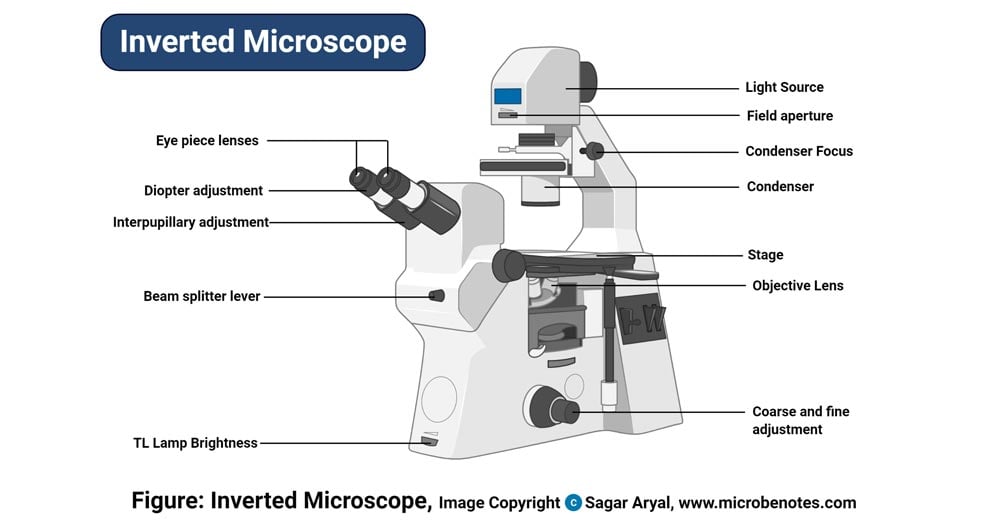
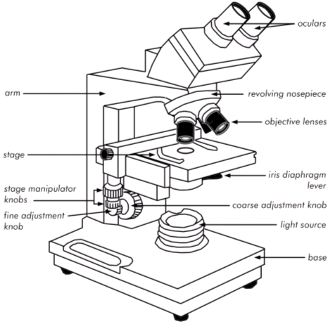
Compound Microscope Parts The monocular (single eye usage) microscope does not need a diopter. Binocular microscopes also swivel (Interpupillary Adjustment) to allow for different distances between the eyes of different individuals. Objective Lenses are the primary optical lenses on a microscope. They range from 4x-100x and typically, include, three, four or five on ... Microscope Parts and Functions Body tube (Head): The body tube connects the eyepiece to the objective lenses. Arm: The arm connects the body tube to the base of the microscope. Coarse adjustment: Brings the specimen into general focus. Fine adjustment: Fine tunes the focus and increases the detail of the specimen. Nosepiece: A rotating turret that houses the objective lenses. Untitled Document [ ] Back to Microscopy. Back to Bio 206
Draw and label a binocular microscope. Find Jobs in Germany: Job Search - Expatica Germany Browse our listings to find jobs in Germany for expats, including jobs for English speakers or those in your native language. microscope worksheet label Binocular Microscope Drawing at GetDrawings | Free download. 16 Pictures about Binocular Microscope Drawing at GetDrawings | Free download : microscope labeling worksheet - Google Search | Science skills, Science, Compound Microscope Diagram Worksheet Answers | Microscope parts and also Color The Microscope Parts Worksheet Answers - Micropedia. A Study of the Microscope and its Functions With a Labeled Diagram The camera present within the microscope captures images to reveal the finer details of the specimen. This microscope can zoom and view the density of a specimen until it is only a micrometer thick and has a magnification ranging between 1,000 - 250,000x on the fluorescent screen. This microscope needs a computer software to yield precise ... Compound Microscope - Diagram (Parts labelled), Principle and Uses What are the 13 parts of a microscope? 1. Eyepiece 2. Eyepiece Tube 3. Objective Lens 4. Stage 5. Stage Clips 6. Nosepiece 7. Fine and Coarse Focus knobs 8. Illuminator 9. Aperture 10. Iris Diaphragm 11. Condenser 12. Condenser Focus Knob 13. The Rack stop Q 5. What are the 11 parts of a compound microscope?
Wild Time 2.3.2015 scale, binoculars, microscope) Science journal supplies Lesson Tips: • We piloted this curriculum with after-school groups where the students didn’t already know each other well. We recommend using the ice breaker Sanctuary Bridge Building to start off the lesson. Science Journals: (20 minutes) 1. Explain that scientists record their observations in journals. We are also scientists … Labeling the Parts of the Microscope | Microscope World Resources Labeling the Parts of the Microscope This activity has been designed for use in homes and schools. Each microscope layout (both blank and the version with answers) are available as PDF downloads. You can view a more in-depth review of each part of the microscope here. Download the Label the Parts of the Microscope PDF printable version here. Labelled Diagram of Compound Microscope The below mentioned article provides a labelled diagram of compound microscope. Part # 1. The Stand: The stand is made up of a heavy foot which carries a curved inclinable limb or arm bearing the body tube. The foot is generally horse shoe-shaped structure (Fig. 2) which rests on table top or any other surface on which the microscope in kept. Compound Microscope Parts - Labeled Diagram and their Functions Numerical Aperture (NA) determines the limit of the Resolution that your microscope can achieve. The value of NA ranges from 0.025 for very low magnification objectives (1x to 4x) to as much as 1.6 for high-performance objectives utilizing specialized immersion oils. The higher the NA, the better the Resolution is. Nosepiece
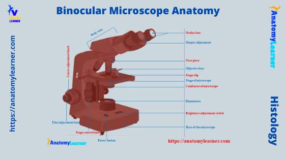
Binocular Microscope Anatomy - AnatomyLearner Now, let's see what the important parts that you should know from the light binocular compound microscope anatomy are - Based on the microscope, Light switch of the microscope, Brightness adjustment switch, A condenser of the microscope, The illuminator of the microscope, Coarse and fine adjustment knobs, Stage control knobs of the microscope, Solved Draw, label and mention function of parts of | Chegg.com Discuss how to use binocular microscope before, during and after. Question : Draw, label and mention function of parts of Binocular Microscope. This problem has been solved! Label the microscope — Science Learning Hub All microscopes share features in common. In this interactive, you can label the different parts of a microscope. Use this with the Microscope parts activity to help students identify and label the main parts of a microscope and then describe their functions. Drag and drop the text labels onto the microscope diagram. DePaul University | DePaul University, Chicago Our Commitment to Anti-Discrimination. DePaul University does not discriminate on the basis of race, color, ethnicity, religion, sex, gender, gender identity, sexual orientation, national origin, age, marital status, pregnancy, parental status, family relationship status, physical or mental disability, military status, genetic information or other status protected by local, state or federal ...
Lifestyle | Daily Life | News | The Sydney Morning Herald The latest Lifestyle | Daily Life news, tips, opinion and advice from The Sydney Morning Herald covering life and relationships, beauty, fashion, health & wellbeing
Fox Files | Fox News Jan 31, 2022 · FOX FILES combines in-depth news reporting from a variety of Fox News on-air talent. The program will feature the breadth, power and journalism of rotating Fox News anchors, reporters and producers.
Compound Microscope: Definition, Diagram, Parts, Uses, Working ... - BYJUS A microscope with a high resolution and uses two sets of lenses providing a 2-dimensional image of the sample. The term compound refers to the usage of more than one lens in the microscope. Also, the compound microscope is one of the types of optical microscopes. The other type of optical microscope is a simple microscope.
Microscope Labeling Diagram | Quizlet Coarse Focus Knob Moves the stage large distances to roughly focus the image. Fine Focus Knob Moves the stage tiny distances to slightly adjust and fine-tune the image focus. Arm Supports the body tube. Objective Lenses Focus and magnify light in differing amounts to view the specimen. Stage Clips Hold the slide in place on the stage. Nosepiece
Parts of a microscope with functions and labeled diagram - Microbe Notes The eyepiece, also known as the ocular is the part used to look through the microscope. Its found at the top of the microscope. Its standard magnification is 10x with an optional eyepiece having magnifications from 5X - 30X. Objective Lens are the major lenses used for specimen visualization. They have a magnification power of 40x-100x.
1910.1001 App B - Detailed Procedures for Asbestos Sampling … Phase contrast microscope with binocular or trinocular head. 6.2.2. Widefield or Huygenian 10X eyepieces (Note: The eyepiece containing the graticule must be a focusing eyepiece. Use a 40X phase objective with a numerical aperture of 0.65 to 0.75). 6.2.3. Kohler illumination (if possible) with green or blue filter. 6.2.4. Walton-Beckett ...
Microscope Parts, Function, & Labeled Diagram - slidingmotion Diaphragm. The diaphragm is also called as iris. This iris situates below the stage of the microscope. The function of the diaphragm is to control the amount of light that focuses on the specimen. This diaphragm can adjust the amount of light and intensity of light that falls on the specimen. In some standard and high-quality microscopes, this ...
Parts of the Microscope with Labeling (also Free Printouts) A microscope is one of the invaluable tools in the laboratory setting. It is used to observe things that cannot be seen by the naked eye. Table of Contents 1. Eyepiece 2. Body tube/Head 3. Turret/Nose piece 4. Objective lenses 5. Knobs (fine and coarse) 6. Stage and stage clips 7. Aperture 9. Condenser 10. Condenser focus knob 11. Iris diaphragm
Drawing Of A Binocular Microscope - Warehouse of Ideas 1) remove the revolving nosepiece and binocular observation tube from the microscope and turn the selector turret on top of the microscope stand to positon "f.c". Source: microspedia.blogspot.com We must create them ourselves. This is the part used to look through the microscope. Source:
39+ Draw And Label A Binocular Microscope Pics Binocular microscope drawing at getdrawings free download. As a compound microscope binocular microscopes use two lenses to magnify the image. Draw a labelled ray diagram of a compound microscope and explain. Feb 12, 2020 · the best free binocular drawing images download from 42 free. This is an online quiz called binocular microscope parts.
9020 QUALITY ASSURANCE/QUALITY CONTROL - Standard Methods for ... 9020 A. Introduction 1. General Considerations Due to the emphasis on microorganisms in water quality standards and enforcement activities and their continuing role in research, process control, and compliance monitoring, laboratories need to implement, document, and effectively operate a quality management system (QS) for microbiological analyses. The QS establishes an environmental testing ...
A Study of the Microscope and its Functions With a Labeled Diagram ... How to Draw Bart Simpson Step By. Christine Walsh. drawing. Microscope Parts. Microscope Slides. Safety Video. Lab Safety. External Lighting. ... Along with a full microscope diagram to label, each part is examined in more detail to properly illustrate each component, its use and function. Click the PREVIEW for a closer look at the PowerPoint ...
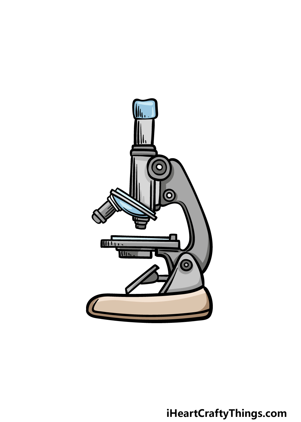
How to draw a microscope step by step beginners guide First, draw two straight lines. You can use a scale for this drawing. Draw lower then draw triazole on top of this drawing. draw a microscope step 1 #Step 2 : Draw same way to next part. draw a microscope step 2 #Step 3 : draw a microscope step 3 #Step 4 : draw a microscope step 4 #Step 5 : draw a microscope step 5 #Step 6 :
Compound Microscope Parts, Functions, and Labeled Diagram Compound Microscope Definitions for Labels. Eyepiece (ocular lens) with or without Pointer: The part that is looked through at the top of the compound microscope. Eyepieces typically have a magnification between 5x & 30x. Monocular or Binocular Head: Structural support that holds & connects the eyepieces to the objective lenses.
China Manufacture of Monocular Microscope Drawing, Monocular Microscope ... China Monocular Microscope Drawing manufacture, a number of high-quality Monocular Microscope Drawing sources of information for you to choose. Inquiry Basket ( 0) ... Multipurpose binocular infinite biology microscope. Monocular Microscope With Low Position Coaxial Coarse. Students Using Monocular Biological Microscope.
How To Draw A Microscope - YouTube Today, we're learning how to draw a cool microscope!👩🎨 JOIN OUR ART HUB MEMBERSHIP! VISIT 🎨 VISIT OUR AMAZON ART SUPPLY S...
Givenchy official site Discover all the collections by Givenchy for women, men & kids and browse the maison's history and heritage
Products - tagged "draw and label a binocular microscope" - laboratorydeal Agronomy Lab Equipment analytical lab Equipment Anatomy lab equipment ayurvedic drug testing laboratory ayurvedic pharmacy instruments B pharmacy Equipments ...
Labelled Diagram Of A Light Microscope - GlobalSpec A schematic diagram for the microscope -based label -free microfluidic light scattering cytometer. ... Draw and label a diagram showing a transverse section of the ileum as seen under a light microscope . Isolation of Halobacterium salinarum retrieved directly from halite brine inclusions. The parts labelled on the diagram are: A, power supply ...
Patent Public Search | USPTO Welcome to Patent Public Search. The Patent Public Search tool is a new web-based patent search application that will replace internal legacy search tools PubEast and PubWest and external legacy search tools PatFT and AppFT.
Microscope Parts & Functions - AmScope Base: A microscope is typically composed of a head or body and a base. The base is the support mechanism. Binocular Microscope: A microscope with a head that has two eyepiece lenses. Nowadays, binocular is typically used to refer to compound or high-power microscopes where the two eyepieces view through a single objective lens.
Microscope Drawing Easy with Label - YouTube Microscope Drawing Easy with Label 886 views Apr 13, 2020 In this video I go over a microscope drawing that is easy with label. There is a blank copy at the end of the video to review...
Untitled Document [ ] Back to Microscopy. Back to Bio 206
Microscope Parts and Functions Body tube (Head): The body tube connects the eyepiece to the objective lenses. Arm: The arm connects the body tube to the base of the microscope. Coarse adjustment: Brings the specimen into general focus. Fine adjustment: Fine tunes the focus and increases the detail of the specimen. Nosepiece: A rotating turret that houses the objective lenses.
Compound Microscope Parts The monocular (single eye usage) microscope does not need a diopter. Binocular microscopes also swivel (Interpupillary Adjustment) to allow for different distances between the eyes of different individuals. Objective Lenses are the primary optical lenses on a microscope. They range from 4x-100x and typically, include, three, four or five on ...








![How To Draw A Microscope Step by Step - [12 Easy Phase]](https://easydrawings.net/wp-content/uploads/2021/01/Overview-for-Microscope-drawing.jpg)





Post a Comment for "40 draw and label a binocular microscope"