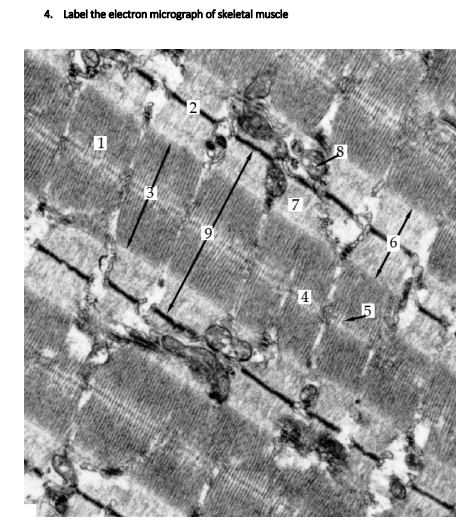
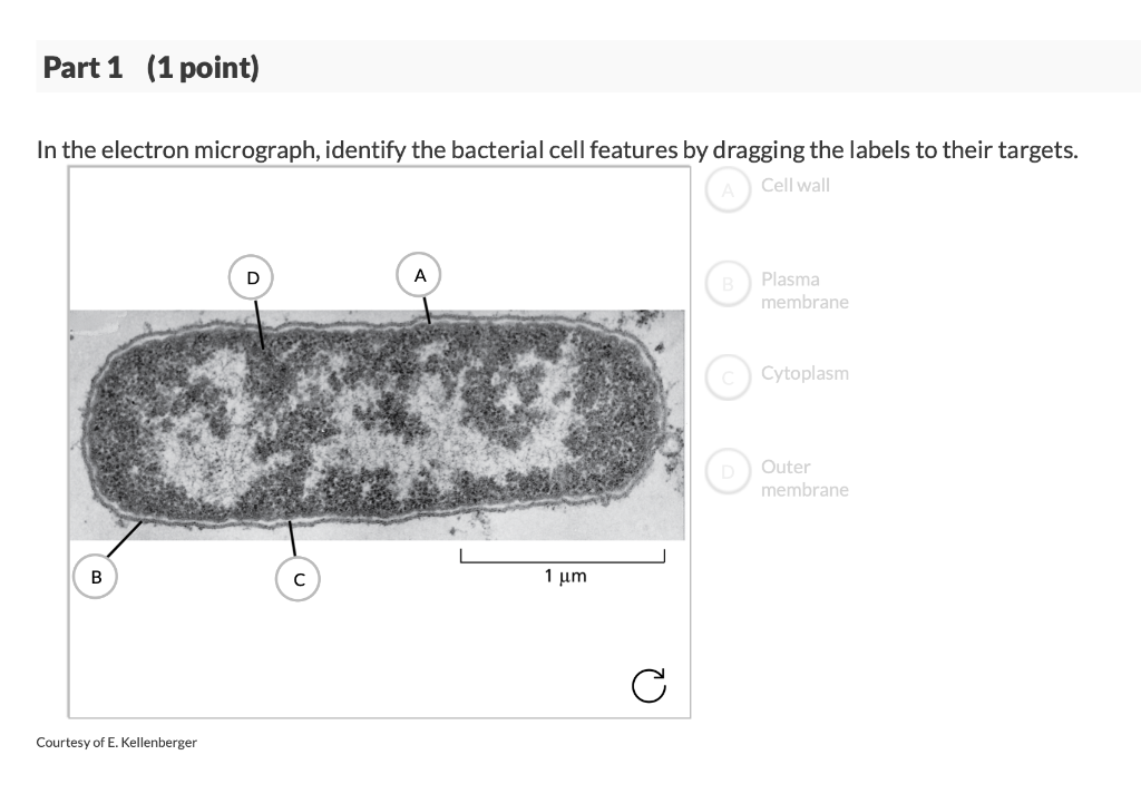
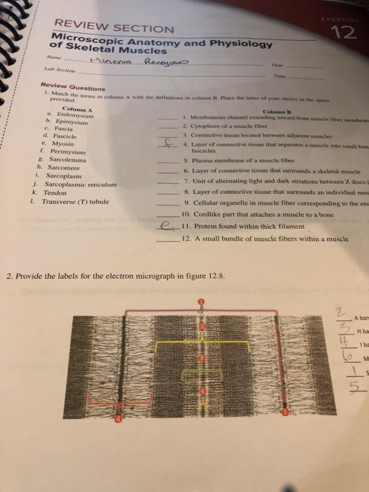
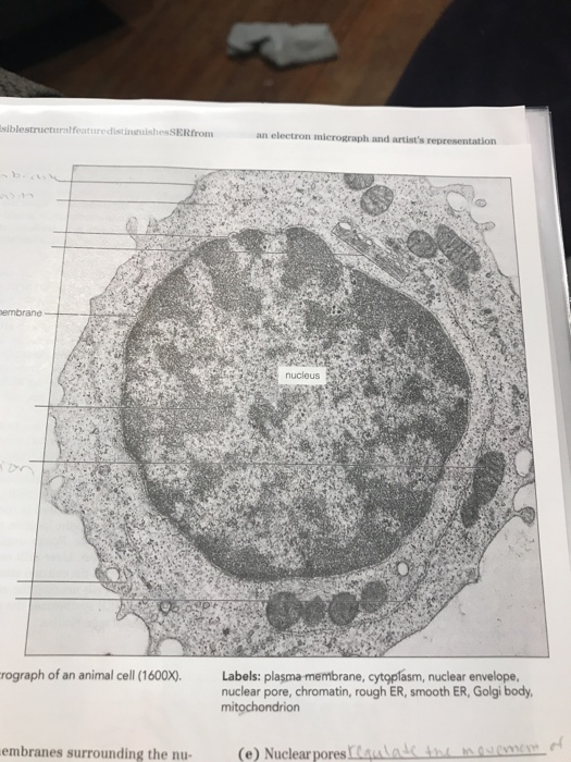
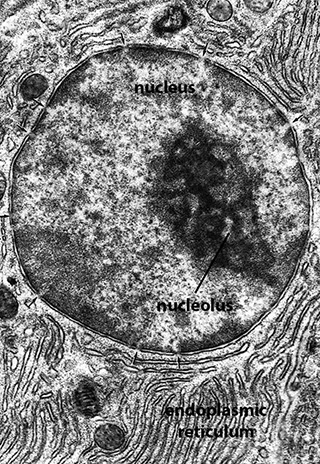
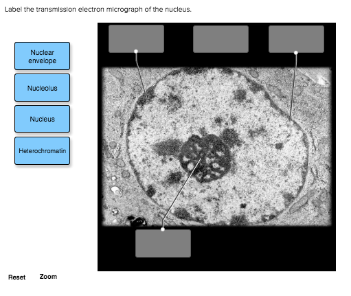
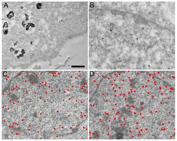
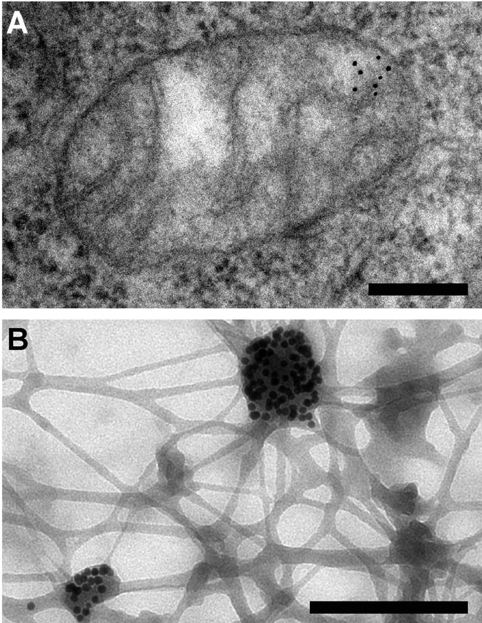
42 provide the labels for the electron micrograph
What is Electron Microscopy? - UMASS Medical School EM images provide key information on the structural basis of cell function and of cell disease. There are two main types of electron microscope – the transmission EM (TEM) and the scanning EM (SEM). The transmission electron microscope is used to view thin specimens (tissue sections, molecules, etc) through which electrons can pass generating ... The Mitochondrion - Molecular Biology of the Cell - NCBI … Mitochondria occupy a substantial portion of the cytoplasmic volume of eucaryotic cells, and they have been essential for the evolution of complex animals. Without mitochondria, present-day animal cells would be dependent on anaerobic glycolysis for all of their ATP. When glucose is converted to pyruvate by glycolysis, only a very small fraction of the total free energy …
Renal System | histology - University of Michigan The close association of arterioles and venules in the vasa recta provide counter-current exchange to help prevent loss of the high electrolyte concentration present in the inner medulla, necessary for the concentration of urine. Capillaries receiving blood from arterioles of the vasa recta are seen throughout the lower medulla. The venules of the vasa recta empty into arcuate …

Provide the labels for the electron micrograph
Ultrasensitive detection of patulin based on a Ag+-driven one … After the formation of stable Au-N bonds between AuNFs and g-C 3 N 4 nanosheets by chemical bonding, the typical transmission electron micrograph of the AuNFs/g-C 3 N 4 composite in Fig. 1 G shows that flower-like particles are fixed on a single nanosheets, indicating that AuNFs were successfully loaded onto the surface of g-C 3 N 4 nanosheets. Stacking Fault - an overview | ScienceDirect Topics Transmission electron micrograph of extended dislocations in Cu–8 at.% Ge solid-solution alloy deformed by 10%. Ge solid-solution alloy deformed by 10%. The stacking fault energy, SFE in short, is an important parameter determining the dissociation of a dislocation, which has a significant influence on the plastic deformation behavior. › topics › materials-scienceStacking Fault - an overview | ScienceDirect Topics Transmission electron micrograph of extended dislocations in Cu–8 at.% Ge solid-solution alloy deformed by 10%. The stacking fault energy, SFE in short, is an important parameter determining the dissociation of a dislocation, which has a significant influence on the plastic deformation behavior.
Provide the labels for the electron micrograph. Cambridge International AS & A Level Biology Coursebook … It is not possible to see an electron beam, so to make the image visible the electron beam has to be projected onto a fluorescent screen. The areas hit by electrons es s-R br am -C screen or photographic film or sensor – shows the image of the specimen Figure 1.16: How a TEM works. Figure 1.15: Scanning electron micrograph (SEM) of a ... Looking at the Structure of Cells in the Microscope A typical animal cell is 10–20 μm in diameter, which is about one-fifth the size of the smallest particle visible to the naked eye. It was not until good light microscopes became available in the early part of the nineteenth century that all plant and animal tissues were discovered to be aggregates of individual cells. This discovery, proposed as the cell doctrine by Schleiden and … Cambridge International AS and A Level Biology ... - Academia.edu BIO1: Maintaining a Balance 1. Most organisms are active in a limited temperature range IDENTIFY THE ROLE OF ENZYMES IN METABOLISM, DESCRIBE THEIR CHEMICAL COMPOSITION AND USE A SIMPLE MODEL TO DESCRIBE … Automated systematic evaluation of cryo-EM specimens with … 23.08.2022 · Cryo-EM density maps of POLG2 collected using SmartScope have been deposited in the Electron Microscopy Data Bank (EMDB) with accession code EMD-25764. Square and hole images and corresponding labels used for training the ML models are available at 10.5281/zenodo.6814642 (square finder) and 10.5281/zenodo.6814652 (hole finder).
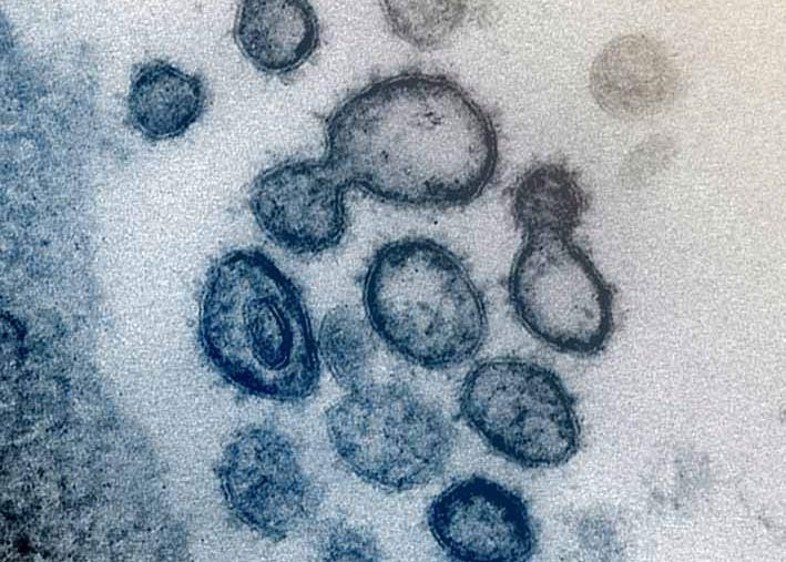
Electron microscope - Wikipedia An electron microscope is a microscope that uses a beam of accelerated electrons as a source of illumination. As the wavelength of an electron can be up to 100,000 times shorter than that of visible light photons, electron microscopes have a higher resolving power than light microscopes and can reveal the structure of smaller objects.. Electron microscopes use shaped magnetic … › cemf › whatisemWhat is Electron Microscopy? - UMASS Medical School Electron microscopy is used in conjunction with a variety of ancillary techniques (e.g. thin sectioning, immuno-labeling, negative staining) to answer specific questions. EM images provide key information on the structural basis of cell function and of cell disease. › topics › materials-scienceStacking Fault - an overview | ScienceDirect Topics Transmission electron micrograph of extended dislocations in Cu–8 at.% Ge solid-solution alloy deformed by 10%. The stacking fault energy, SFE in short, is an important parameter determining the dissociation of a dislocation, which has a significant influence on the plastic deformation behavior. Stacking Fault - an overview | ScienceDirect Topics Transmission electron micrograph of extended dislocations in Cu–8 at.% Ge solid-solution alloy deformed by 10%. Ge solid-solution alloy deformed by 10%. The stacking fault energy, SFE in short, is an important parameter determining the dissociation of a dislocation, which has a significant influence on the plastic deformation behavior.
Ultrasensitive detection of patulin based on a Ag+-driven one … After the formation of stable Au-N bonds between AuNFs and g-C 3 N 4 nanosheets by chemical bonding, the typical transmission electron micrograph of the AuNFs/g-C 3 N 4 composite in Fig. 1 G shows that flower-like particles are fixed on a single nanosheets, indicating that AuNFs were successfully loaded onto the surface of g-C 3 N 4 nanosheets.













Post a Comment for "42 provide the labels for the electron micrograph"